Departments of Rehabilitation, Ruijin Hospital, School of Medicine, Shanghai Jiao Tong University, Shanghai 200025, China.
Med-X Research Institute and School of Biomedical Engineering, Shanghai Jiao Tong University, Shanghai 200030, China.
Theranostics. 2019 Jul 9;9(17):4923-4934. doi: 10.7150/thno.32676. eCollection 2019.
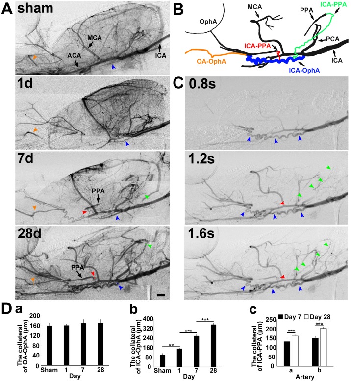
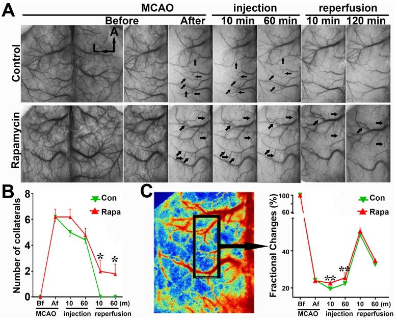
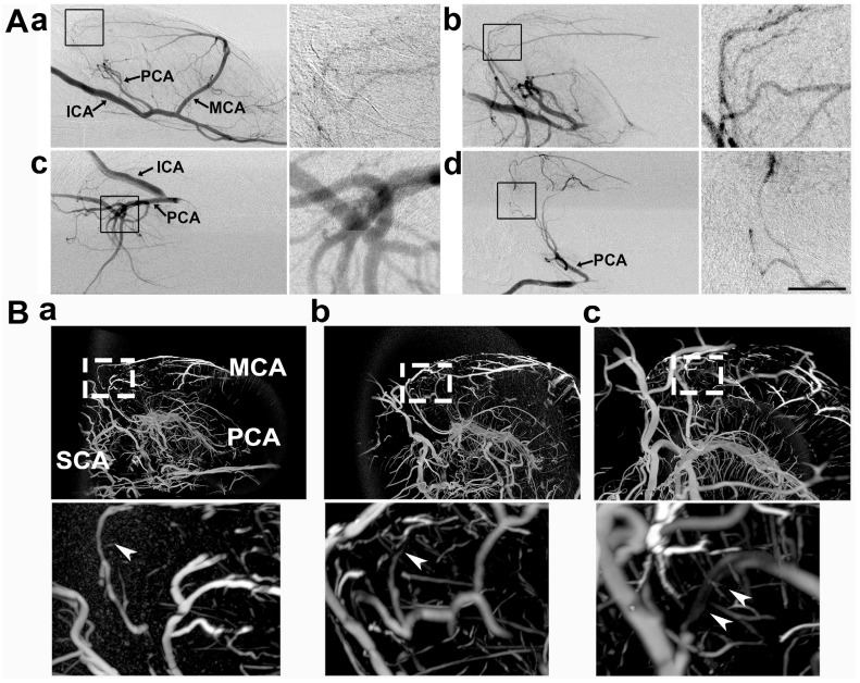
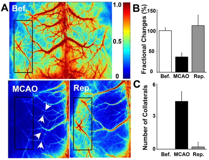
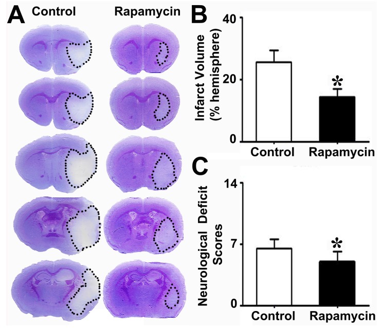
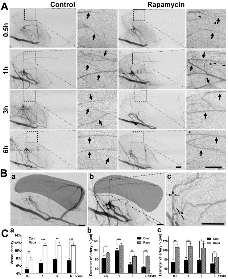
: Brain collaterals contribute to improving ischemic stroke outcomes. However, dynamic and timely investigations of collateral blood flow and collateral restoration in whole brains of living animals have rarely been reported. : Using multiple modalities of imaging, including synchrotron radiation angiography, laser speckle imaging, and micro-CT imaging, we dynamically explored collateral circulation throughout the whole brain in the rodent middle cerebral artery occlusion model. : We demonstrated that compared to control animals, 4 neocollaterals gradually formed between the intra- and extra-arteries in the skull base of model animals after occlusion (<0.05). Two main collaterals were critical to the supply of blood from the posterior to the middle cerebral artery territory in the deep brain (<0.05). Abundant small vessel and capillary anastomoses were detected on the surface of the cortex between the posterior and middle cerebral artery and between the anterior and middle cerebral artery (<0.05). Collateral perfusion occurred immediately (≈15 min) and was maintained for up to 14 days after occlusion. Further study revealed that administration of rapamycin at 15 min after MCAO dilated the existing collateral vessels and promoted collateral perfusion. : Our results provide evidence of collateral functional perfusion in the skull base, deep brain, and surface of the cortex. Rapamycin was capable of enlarging the diameter of collaterals, potentially extending the time window for ischemic stroke therapy.
脑侧支循环有助于改善缺血性脑卒中的预后。然而,活体动物全脑侧支血流和侧支恢复的动态和实时研究很少报道。
使用多种成像模式,包括同步辐射血管造影、激光散斑成像和 micro-CT 成像,我们在啮齿动物大脑中动脉闭塞模型中动态研究了整个大脑的侧支循环。
我们证明,与对照动物相比,闭塞后模型动物颅底的 intra- 和 extra- 动脉之间逐渐形成了 4 条新的侧支(<0.05)。2 条主要侧支对大脑深部分支从中动脉到大脑中动脉供血至关重要(<0.05)。在大脑中动脉和大脑前动脉之间的大脑表面检测到丰富的小血管和毛细血管吻合(<0.05)。侧支灌注在闭塞后立即发生(≈15 min),并持续 14 天。进一步的研究表明,在 MCAO 后 15 分钟给予雷帕霉素可扩张现有的侧支血管并促进侧支灌注。
我们的研究结果提供了颅底、大脑深部和大脑表面侧支功能性灌注的证据。雷帕霉素能够扩大侧支的直径,可能延长缺血性脑卒中治疗的时间窗。