Department of Drug Discovery & Biomedical Sciences, Medical University of South Carolina , Charleston , SC , USA.
Department of Pediatrics, Medical University of South Carolina , Charleston , SC , USA.
Int J Hyperthermia. 2019;36(1):817-826. doi: 10.1080/02656736.2019.1642521.
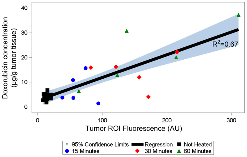
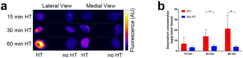
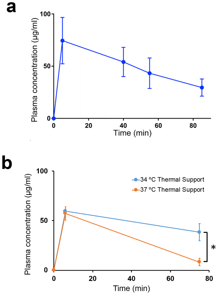
Thermosensitive liposomal doxorubicin (TSL-Dox) is a promising stimuli-responsive nanoparticle drug delivery system that rapidly releases the contained drug in response to hyperthermia (HT) (>40 °C). Combined with localized heating, TSL-Dox allows highly localized delivery. The goals of this study were to demonstrate that real-time fluorescence imaging can visualize drug uptake during delivery, and can predict tumor drug uptake. Nude mice carrying subcutaneous tumors (Lewis lung carcinoma) were anesthetized and injected with TSL-Dox (5 mg/kg dose). Localized HT was induced by heating tumors for 15, 30 or 60 min via a custom-designed HT probe placed superficially at the tumor location. fluorescence imaging (excitation 523 nm, emission 610 nm) was performed before, during, and for 5 min following HT. After imaging, tumors were extracted, drug uptake was quantified by high-performance liquid chromatography, and correlated with fluorescence. Plasma samples were obtained before and after HT to measure TSL-Dox pharmacokinetics. Local drug uptake could be visualized in real-time during HT. Compared to unheated control tumors, fluorescence of heated tumors increased by 4.6-fold (15 min HT), 9.3-fold (30 min HT), and 13.2-fold (60 min HT). HT duration predicted tumor drug uptake ( = .02), with tumor drug concentrations of 4.2 ± 1.3 µg/g (no HT), 7.1 ± 5.9 µg/g (15 min HT), 14.1 ± 6.7 µg/g (30 min HT) and 21.4 ± 12.6 µg/g (60 min HT). There was good correlation ( = 0.67) between fluorescence of the tumor region and tumor drug uptake. Real-time fluorescence imaging can visualize drug uptake during delivery, and can predict tumor drug uptake.
热敏脂质体阿霉素(TSL-Dox)是一种有前途的刺激响应纳米颗粒药物递送系统,它可以在高温(>40°C)下迅速释放包含的药物。与局部加热相结合,TSL-Dox 允许高度局部递送。本研究的目的是证明实时荧光成像可以可视化药物在递送过程中的摄取,并可以预测肿瘤药物摄取。携带皮下肿瘤(Lewis 肺癌)的裸鼠在麻醉下,以 5mg/kg 剂量注射 TSL-Dox。通过将定制设计的 HT 探头放置在肿瘤位置的表面来诱导局部 HT15、30 或 60 分钟。在 HT 之前、期间和之后进行 5 分钟的荧光成像(激发 523nm,发射 610nm)。成像后,提取肿瘤,通过高效液相色谱法定量测定药物摄取,并与荧光相关。在 HT 前后获得血浆样本以测量 TSL-Dox 药代动力学。在 HT 期间可以实时可视化局部药物摄取。与未加热的对照肿瘤相比,加热肿瘤的荧光增加了 4.6 倍(15 分钟 HT),9.3 倍(30 分钟 HT)和 13.2 倍(60 分钟 HT)。HT 持续时间预测肿瘤药物摄取( = .02),无 HT 时肿瘤药物浓度为 4.2 ± 1.3 μg/g,15 分钟 HT 时为 7.1 ± 5.9 μg/g,30 分钟 HT 时为 14.1 ± 6.7 μg/g,60 分钟 HT 时为 21.4 ± 12.6 μg/g。肿瘤区域的荧光与肿瘤药物摄取之间存在良好的相关性( = 0.67)。实时荧光成像可以可视化药物在递送过程中的摄取,并可以预测肿瘤药物摄取。