• 文献检索
  • 文档翻译
  • 深度研究
  • 学术资讯
  • Suppr Zotero 插件Zotero 插件
  • 邀请有礼
  • 套餐&价格
  • 历史记录
应用&插件
Suppr Zotero 插件Zotero 插件浏览器插件Mac 客户端Windows 客户端微信小程序
定价
高级版会员购买积分包购买API积分包
服务
文献检索文档翻译深度研究API 文档MCP 服务
关于我们
关于 Suppr公司介绍联系我们用户协议隐私条款
关注我们

Suppr 超能文献

核心技术专利:CN118964589B侵权必究
粤ICP备2023148730 号-1Suppr @ 2026

文献检索

告别复杂PubMed语法,用中文像聊天一样搜索,搜遍4000万医学文献。AI智能推荐,让科研检索更轻松。

立即免费搜索

文件翻译

保留排版,准确专业,支持PDF/Word/PPT等文件格式,支持 12+语言互译。

免费翻译文档

深度研究

AI帮你快速写综述,25分钟生成高质量综述,智能提取关键信息,辅助科研写作。

立即免费体验

光感受器神经节细胞在出生后发育过程中的细胞特性。

Cellular properties of intrinsically photosensitive retinal ganglion cells during postnatal development.

机构信息

Department of Neurobiology, Northwestern University, Evanston, IL, USA.

出版信息

Neural Dev. 2019 Aug 30;14(1):8. doi: 10.1186/s13064-019-0132-2.

DOI:10.1186/s13064-019-0132-2
PMID:31470901
原文链接:https://pmc.ncbi.nlm.nih.gov/articles/PMC6716945/
Abstract

BACKGROUND

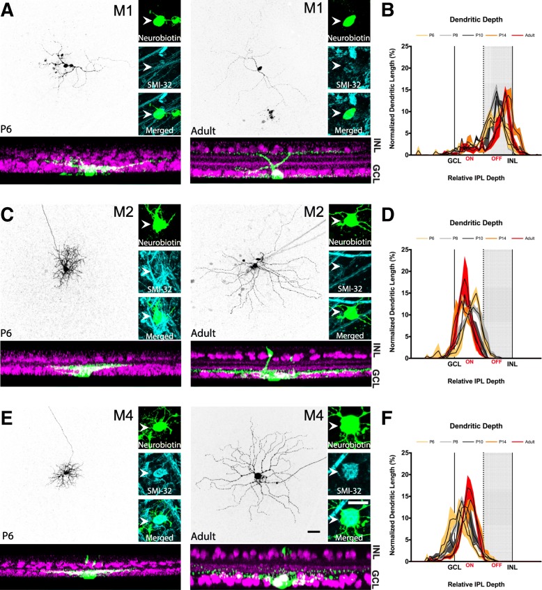
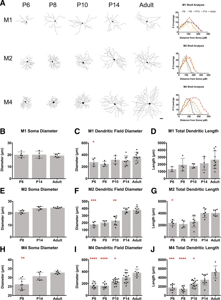
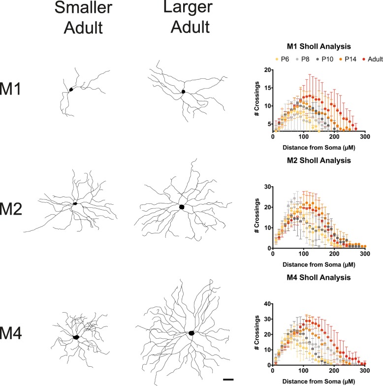
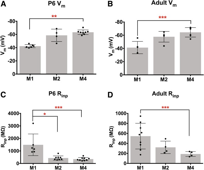
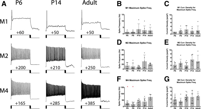
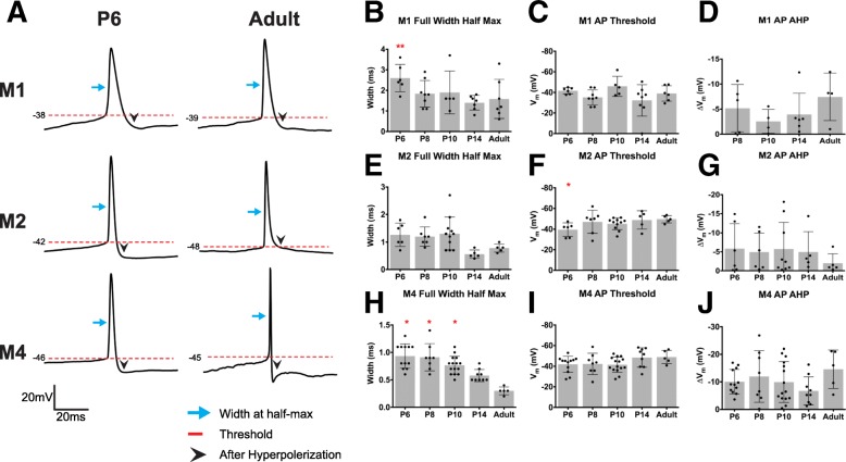
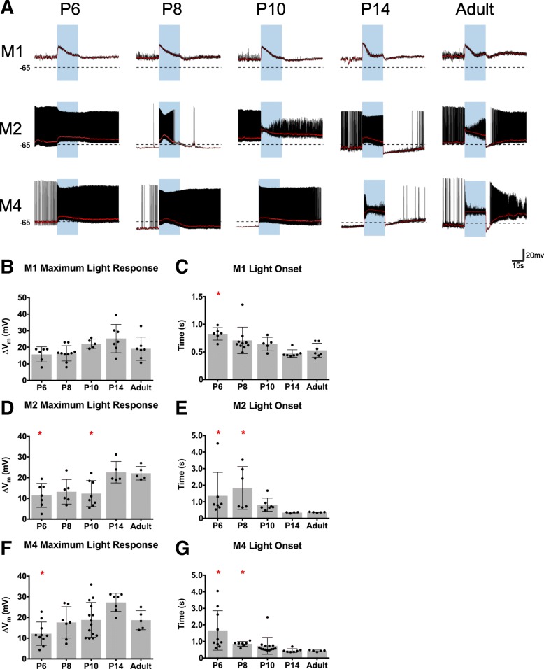
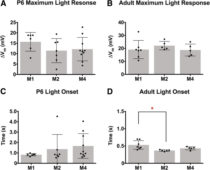
Melanopsin-expressing, intrinsically photosensitive retinal ganglion cells (ipRGCs) respond directly to light and have been shown to mediate a broad variety of visual behaviors in adult animals. ipRGCs are also the first light sensitive cells in the developing retina, and have been implicated in a number of retinal developmental processes such as pruning of retinal vasculature and refinement of retinofugal projections. However, little is currently known about the properties of the six ipRGC subtypes during development, and how these cells act to influence retinal development. We therefore sought to characterize the structure, physiology, and birthdate of the most abundant ipRGC subtypes, M1, M2, and M4, at discrete postnatal developmental timepoints.

METHODS

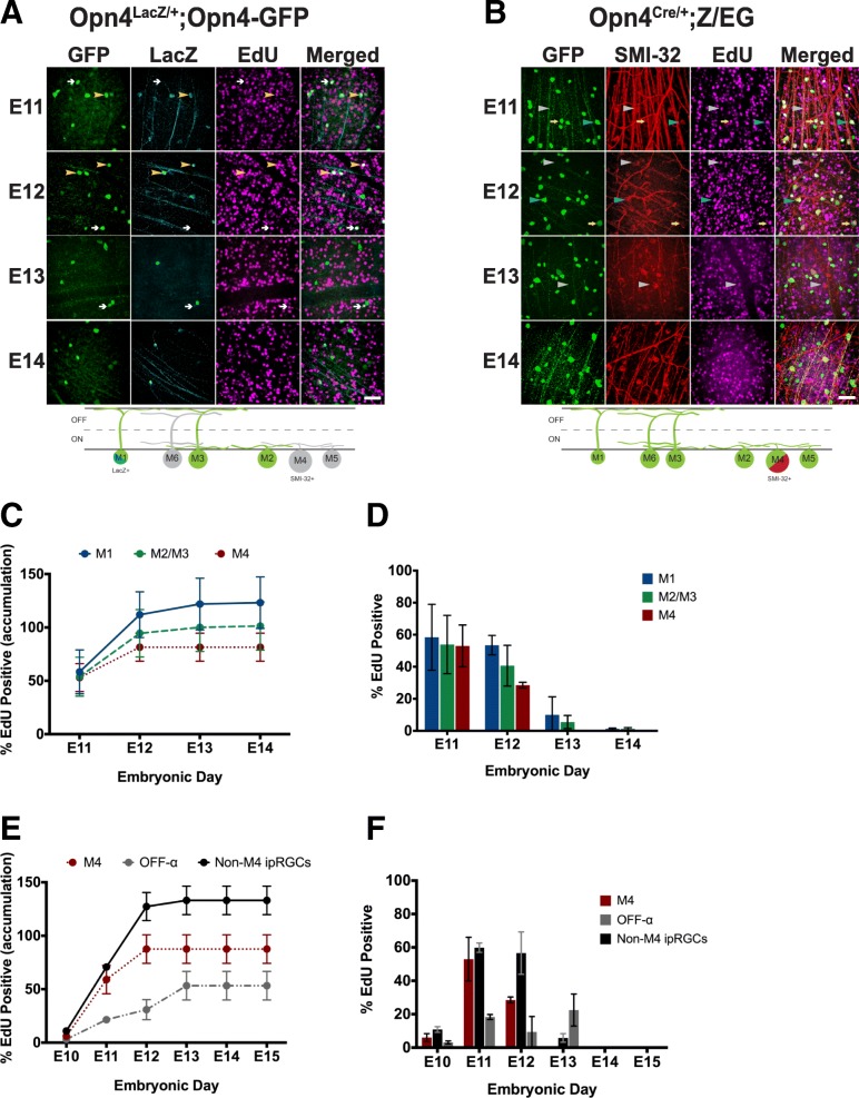
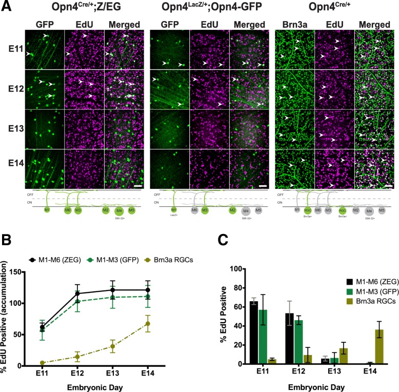
We utilized whole cell patch clamp to measure the electrophysiological and morphological properties of ipRGC subtypes through postnatal development. We also used EdU labeling to determine the embryonic timepoints at which ipRGC subtypes terminally differentiate.

RESULTS

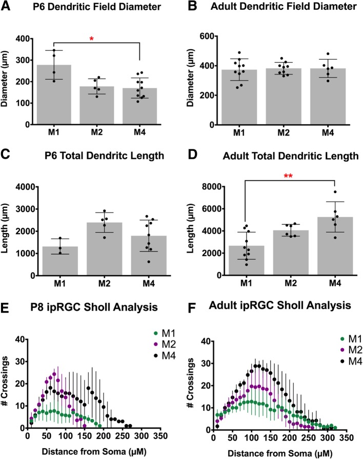
Our data show that ipRGC subtypes are distinguishable from each other early in postnatal development. Additionally, we find that while ipRGC subtypes terminally differentiate at similar embryonic stages, the subtypes reach adult-like morphology and physiology at different developmental timepoints.

CONCLUSIONS

This work provides a broad assessment of ipRGC morphological and physiological properties during the postnatal stages at which they are most influential in modulating retinal development, and lays the groundwork for further understanding of the specific role of each ipRGC subtype in influencing retinal and visual system development.

摘要

背景

表达黑视蛋白的光敏感视网膜神经节细胞(ipRGC)直接对光起反应,并已被证明在成年动物中介导广泛的各种视觉行为。ipRGC 也是发育中视网膜中的第一光敏感细胞,并且已经涉及到许多视网膜发育过程,如视网膜血管系统的修剪和视网膜投射的细化。然而,目前对于发育过程中六种 ipRGC 亚型的特性以及这些细胞如何影响视网膜发育知之甚少。因此,我们试图在离散的出生后发育时间点表征最丰富的 ipRGC 亚型 M1、M2 和 M4 的结构、生理学和出生日期。

方法

我们利用全细胞膜片钳技术测量 ipRGC 亚型在出生后的发育过程中的电生理和形态特性。我们还使用 EdU 标记来确定 ipRGC 亚型终末分化的胚胎时间点。

结果

我们的数据表明,ipRGC 亚型在出生后的早期就可以彼此区分开来。此外,我们发现虽然 ipRGC 亚型在相似的胚胎阶段终末分化,但它们达到成人样形态和生理学的时间点不同。

结论

这项工作提供了在最能调节视网膜发育的出生后阶段对 ipRGC 形态和生理学特性的广泛评估,并为进一步了解每个 ipRGC 亚型在影响视网膜和视觉系统发育中的具体作用奠定了基础。

https://cdn.ncbi.nlm.nih.gov/pmc/blobs/eb0e/6716945/630bc9168143/13064_2019_132_Fig12_HTML.jpg
https://cdn.ncbi.nlm.nih.gov/pmc/blobs/eb0e/6716945/7be7b92691f9/13064_2019_132_Fig1_HTML.jpg
https://cdn.ncbi.nlm.nih.gov/pmc/blobs/eb0e/6716945/bb6ed0e70a34/13064_2019_132_Fig2_HTML.jpg
https://cdn.ncbi.nlm.nih.gov/pmc/blobs/eb0e/6716945/f0cd374a6724/13064_2019_132_Fig3_HTML.jpg
https://cdn.ncbi.nlm.nih.gov/pmc/blobs/eb0e/6716945/af5aae826642/13064_2019_132_Fig4_HTML.jpg
https://cdn.ncbi.nlm.nih.gov/pmc/blobs/eb0e/6716945/23aec5a64e27/13064_2019_132_Fig5_HTML.jpg
https://cdn.ncbi.nlm.nih.gov/pmc/blobs/eb0e/6716945/1c918e648c88/13064_2019_132_Fig6_HTML.jpg
https://cdn.ncbi.nlm.nih.gov/pmc/blobs/eb0e/6716945/b44143a23d9d/13064_2019_132_Fig7_HTML.jpg
https://cdn.ncbi.nlm.nih.gov/pmc/blobs/eb0e/6716945/726767f306a8/13064_2019_132_Fig8_HTML.jpg
https://cdn.ncbi.nlm.nih.gov/pmc/blobs/eb0e/6716945/c3f00ffd5b4a/13064_2019_132_Fig9_HTML.jpg
https://cdn.ncbi.nlm.nih.gov/pmc/blobs/eb0e/6716945/b149b3592e8b/13064_2019_132_Fig10_HTML.jpg
https://cdn.ncbi.nlm.nih.gov/pmc/blobs/eb0e/6716945/1e52e0596d66/13064_2019_132_Fig11_HTML.jpg
https://cdn.ncbi.nlm.nih.gov/pmc/blobs/eb0e/6716945/630bc9168143/13064_2019_132_Fig12_HTML.jpg

相似文献

1
Cellular properties of intrinsically photosensitive retinal ganglion cells during postnatal development.光感受器神经节细胞在出生后发育过程中的细胞特性。
Neural Dev. 2019 Aug 30;14(1):8. doi: 10.1186/s13064-019-0132-2.
2
Photoreceptive Ganglion Cells Drive Circuits for Local Inhibition in the Mouse Retina.感光神经节细胞驱动小鼠视网膜中的局部抑制回路。
J Neurosci. 2021 Feb 17;41(7):1489-1504. doi: 10.1523/JNEUROSCI.0674-20.2020. Epub 2021 Jan 4.
3
M1 ipRGCs Influence Visual Function through Retrograde Signaling in the Retina.M1型内在光敏视网膜神经节细胞通过视网膜中的逆行信号传导影响视觉功能。
J Neurosci. 2016 Jul 6;36(27):7184-97. doi: 10.1523/JNEUROSCI.3500-15.2016.
4
Morphological Identification of Melanopsin-Expressing Retinal Ganglion Cell Subtypes in Mice.小鼠中表达黑视蛋白的视网膜神经节细胞亚型的形态学鉴定
Methods Mol Biol. 2018;1753:275-287. doi: 10.1007/978-1-4939-7720-8_19.
5
Type I intrinsically photosensitive retinal ganglion cells of early post-natal development correspond to the M4 subtype.出生后早期发育阶段的I型内在光敏性视网膜神经节细胞对应于M4亚型。
Neural Dev. 2015 Jun 21;10:17. doi: 10.1186/s13064-015-0042-x.
6
Retinal Waves Modulate an Intraretinal Circuit of Intrinsically Photosensitive Retinal Ganglion Cells.视网膜波调节内在光敏性视网膜神经节细胞的视网膜内回路。
J Neurosci. 2016 Jun 29;36(26):6892-905. doi: 10.1523/JNEUROSCI.0572-16.2016.
7
Immunotoxin-induced ablation of melanopsin retinal ganglion cells in a non-murine mammalian model.免疫毒素诱导的非鼠类哺乳动物模型中黑视蛋白视网膜神经节细胞的消融。
J Comp Neurol. 2009 Sep 10;516(2):125-40. doi: 10.1002/cne.22103.
8
Physiologic diversity and development of intrinsically photosensitive retinal ganglion cells.内在光敏性视网膜神经节细胞的生理多样性与发育
Neuron. 2005 Dec 22;48(6):987-99. doi: 10.1016/j.neuron.2005.09.031.
9
Structure and function of bistratified intrinsically photosensitive retinal ganglion cells in the mouse.双分层内在感光视网膜神经节细胞在小鼠中的结构与功能。
J Comp Neurol. 2011 Jun 1;519(8):1492-504. doi: 10.1002/cne.22579.
10
Intrinsic photosensitive retinal ganglion cells in the diurnal rodent, Arvicanthis ansorgei.昼行性鼠类非洲沙鼠中的内在光敏感视网膜神经节细胞。
PLoS One. 2013 Aug 9;8(8):e73343. doi: 10.1371/journal.pone.0073343. eCollection 2013.

引用本文的文献

1
Protocol for the purification and culture of primary retinal ganglion cells and development of common pathological models.原代视网膜神经节细胞的纯化培养及常见病理模型构建方案。
J Mol Histol. 2025 Aug 8;56(4):260. doi: 10.1007/s10735-025-10536-x.
2
Atypical retinal ganglion cell function in a mouse model of Fragile X syndrome.脆性X综合征小鼠模型中的非典型视网膜神经节细胞功能
J Neurosci. 2025 Jun 4. doi: 10.1523/JNEUROSCI.0013-25.2025.
3
Two-photon activation, deactivation, and coherent control of melanopsin in live cells.活细胞中黑视蛋白的双光子激活、失活及相干控制。

本文引用的文献

1
Light Affects Mood and Learning through Distinct Retina-Brain Pathways.光通过独特的视网膜-大脑通路影响情绪和学习。
Cell. 2018 Sep 20;175(1):71-84.e18. doi: 10.1016/j.cell.2018.08.004. Epub 2018 Aug 30.
2
Melanopsin Phototransduction Is Repurposed by ipRGC Subtypes to Shape the Function of Distinct Visual Circuits.黑视蛋白光转导被 ipRGC 亚型重新用于塑造不同视觉回路的功能。
Neuron. 2018 Aug 22;99(4):754-767.e4. doi: 10.1016/j.neuron.2018.06.032. Epub 2018 Jul 12.
3
Biophysical Variation within the M1 Type of Ganglion Cell Photoreceptor.
bioRxiv. 2025 Mar 30:2025.03.26.645437. doi: 10.1101/2025.03.26.645437.
4
Light activation of the dopaminergic system occurs after eye-opening in the mouse retina.在小鼠视网膜中,多巴胺能系统的光激活发生在睁眼之后。
Front Ophthalmol (Lausanne). 2023 May 9;3:1184627. doi: 10.3389/fopht.2023.1184627. eCollection 2023.
5
Atypical retinal function in a mouse model of Fragile X syndrome.脆性X综合征小鼠模型中的非典型视网膜功能。
bioRxiv. 2024 Mar 17:2024.03.15.585283. doi: 10.1101/2024.03.15.585283.
6
Assessing perceptual chromatic equiluminance using a reflexive pupillary response.利用瞳孔反射评估知觉等色明度。
Sci Rep. 2024 Jan 29;14(1):2420. doi: 10.1038/s41598-024-51982-z.
7
Melanopsin activates divergent phototransduction pathways in intrinsically photosensitive retinal ganglion cell subtypes.黑视素激活内在光敏感视网膜神经节细胞亚型中的不同光转导途径。
Elife. 2023 Nov 8;12:e80749. doi: 10.7554/eLife.80749.
8
Circuit mechanisms underlying embryonic retinal waves.胚胎视网膜波的电路机制。
Elife. 2023 Feb 15;12:e81983. doi: 10.7554/eLife.81983.
9
The Newborn's Reaction to Light as the Determinant of the Brain's Activation at Human Birth.新生儿对光的反应作为人类出生时大脑激活的决定因素。
Front Integr Neurosci. 2022 Sep 2;16:933426. doi: 10.3389/fnint.2022.933426. eCollection 2022.
10
Morphological Factors that Underlie Neural Sensitivity to Stimulation in the Retina.视网膜中神经对刺激敏感的形态学因素。
Adv Nanobiomed Res. 2021 Dec;1(12). doi: 10.1002/anbr.202100069. Epub 2021 Sep 1.
M1 型神经节细胞光感受器的生物物理变化。
Cell Rep. 2017 Oct 24;21(4):1048-1062. doi: 10.1016/j.celrep.2017.09.095.
4
A Population Representation of Absolute Light Intensity in the Mammalian Retina.哺乳动物视网膜中绝对光强度的群体表征。
Cell. 2017 Nov 2;171(4):865-876.e16. doi: 10.1016/j.cell.2017.09.005. Epub 2017 Sep 28.
5
A subset of ipRGCs regulates both maturation of the circadian clock and segregation of retinogeniculate projections in mice.在小鼠中,一部分 intrinsically photosensitive retinal ganglion cells(ipRGCs)既调节生物钟的成熟,又调节视网膜神经节细胞投射的分离。
Elife. 2017 Jun 15;6:e22861. doi: 10.7554/eLife.22861.
6
Retinal Waves Modulate an Intraretinal Circuit of Intrinsically Photosensitive Retinal Ganglion Cells.视网膜波调节内在光敏性视网膜神经节细胞的视网膜内回路。
J Neurosci. 2016 Jun 29;36(26):6892-905. doi: 10.1523/JNEUROSCI.0572-16.2016.
7
Cardinal Orientation Selectivity Is Represented by Two Distinct Ganglion Cell Types in Mouse Retina.小鼠视网膜中两种不同的神经节细胞类型代表主要方向选择性。
J Neurosci. 2016 Mar 16;36(11):3208-21. doi: 10.1523/JNEUROSCI.4554-15.2016.
8
All spiking, sustained ON displaced amacrine cells receive gap-junction input from melanopsin ganglion cells.所有产生峰电位的、持续开启的移位无长突细胞都从黑视蛋白神经节细胞接收缝隙连接输入。
Curr Biol. 2015 Nov 2;25(21):2763-2773. doi: 10.1016/j.cub.2015.09.018. Epub 2015 Oct 1.
9
Type I intrinsically photosensitive retinal ganglion cells of early post-natal development correspond to the M4 subtype.出生后早期发育阶段的I型内在光敏性视网膜神经节细胞对应于M4亚型。
Neural Dev. 2015 Jun 21;10:17. doi: 10.1186/s13064-015-0042-x.
10
Automated computation of arbor densities: a step toward identifying neuronal cell types.树突密度的自动计算:迈向识别神经元细胞类型的一步。
Front Neuroanat. 2014 Nov 25;8:139. doi: 10.3389/fnana.2014.00139. eCollection 2014.