Russell H. Morgan Department of Radiology and Radiological Science, Division of MR Research, Johns Hopkins University School of Medicine, Baltimore, Maryland.
F.M. Kirby Research Center for Functional Brain Imaging, Kennedy Krieger Institute, Baltimore, Maryland.
Magn Reson Med. 2020 Mar;83(3):1066-1080. doi: 10.1002/mrm.27972. Epub 2019 Sep 4.
To evaluate different T -oxygenation calibrations for estimating venous oxygenation in people with sickle cell anemia (SCA).
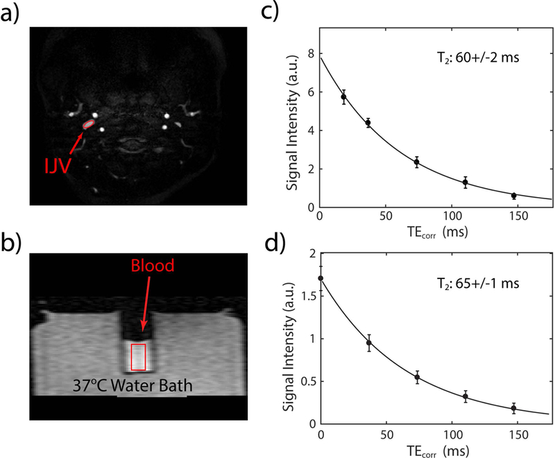
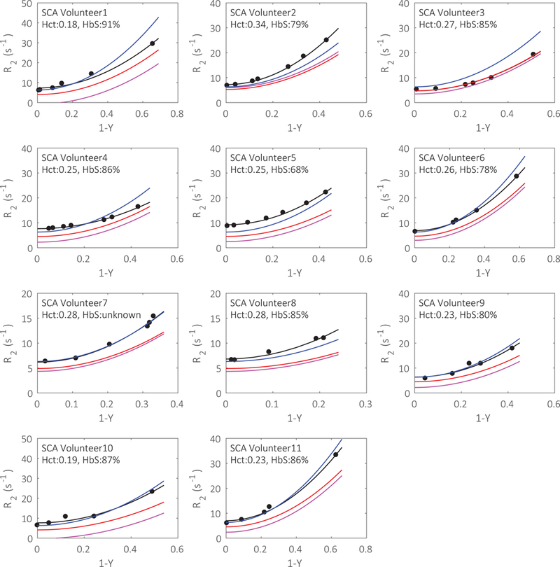
Blood T values were measured at 3 T in the internal jugular veins of 12 healthy volunteers and 11 SCA participants with no history of stroke, recent transfusion, or renal impairment. T -oxygenation relationships of both sickled and normal blood samples were calibrated individually and compared with values generated from published models. After converting venous T values to venous oxygenation, whole-brain oxygen extraction fraction and cerebral metabolic rate of oxygen were calculated.
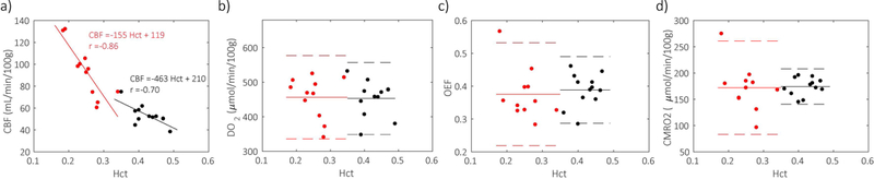
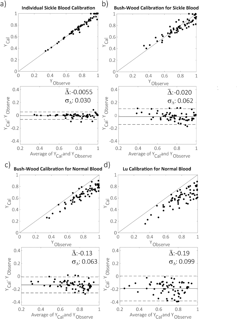
Sickle blood samples' oxygenation values calculated from our individual calibrations agreed well with measurements using a blood analyzer, whereas previous T calibrations based on normal blood samples showed 13%-19% underestimation. Meanwhile, oxygenation values calculated from previous grouped T calibration for sickle blood agreed well with experimental measurement on averaged values, but showed up to 20% variation for several individual samples. Using individual T calibrations, the whole-brain oxygen extraction fraction and cerebral metabolic rate of oxygen of SCA participants were 0.38 ± 0.08 and 172 ± 42 µmol/min/100 g, respectively, which were comparable to those values measured on healthy volunteers.
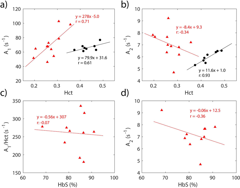
Our results confirm that sickle blood T values not only depend on the hematocrit and oxygenation values, but also on other hematological factors. The individual T calibrations minimized the effect of heterogeneity of sickle blood between different SCA populations and improved the accuracy of T -based oximetry. The measured oxygen extraction fraction and cerebral metabolic rate of oxygen of this group of SCA participants were found to not differ significantly from those of healthy individuals.
评估不同 T- 氧合校准方法在评估镰状细胞贫血(SCA)患者静脉氧合中的应用。
在 3T 下测量 12 名健康志愿者和 11 名无卒中史、近期输血或肾功能不全史的 SCA 参与者颈内静脉的血液 T 值。分别对镰状和正常血液样本的 T- 氧合关系进行校准,并与已发表模型生成的值进行比较。将静脉 T 值转换为静脉氧合后,计算全脑氧摄取分数和脑氧代谢率。
我们的个体校准计算出的镰状血样本的氧合值与血液分析仪的测量值吻合良好,而以前基于正常血样本的 T 校准则低估了 13%-19%。同时,以前基于镰状血分组 T 校准计算出的氧合值与平均测量值吻合良好,但对几个个体样本的差异高达 20%。使用个体 T 校准,SCA 参与者的全脑氧摄取分数和脑氧代谢率分别为 0.38 ± 0.08 和 172 ± 42µmol/min/100g,与健康志愿者的测量值相当。
我们的结果证实,镰状血的 T 值不仅取决于血细胞比容和氧合值,还取决于其他血液学因素。个体 T 校准最大限度地减少了不同 SCA 人群中镰状血之间的异质性的影响,提高了基于 T 的血氧测定的准确性。该组 SCA 参与者的测量氧摄取分数和脑氧代谢率与健康个体无显著差异。