Fulciniti Franco, Cipolletta Campanile Anna, Malzone Maria Gabriella, Chiofalo Maria Grazia, Capiluongo Anna, Monaco Mario, Di Maio Nunzia, Sandomenico Fabio, Botti Gerardo, Chiappetta Gennaro, Vuttariello Emilia, Pezzullo Luciano
Clinical Cytopathology Service, Istituto Cantonale di Patologia, Locarno, Switzerland.
Cytohistopathology Laboratory, Istituto Diagnostico Varelli, Naples, Italy.
Clin Endocrinol (Oxf). 2019 Dec;91(6):851-859. doi: 10.1111/cen.14089. Epub 2019 Oct 1.
Fine needle cytology (FNC) is the first-line diagnostic method to determine the benign or malignant nature of thyroid nodules. The gray zone of cytological classifications remains, however, a crucial and challenging area for cytopathologists.
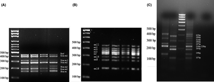
DESIGN, PATIENTS AND MEASUREMENTS: In the present study, 141 thyroid cytological samples, with ultrasonographic suspicious features, have been prospectively analysed. Molecular analyses were performed by an innovative technology using two multiplex PCRs for the amplification of BRAF, N-H-K-RAS and RET exon genes. RNA samples were studied for RET/PTC1 and RET/PTC3 rearrangements by PCR amplification, and the conditions were set-up to study, with a single experiment, both wild-type PAX8 and PAX8/PPARɣ rearrangements. In total, 111 samples were examined for BRAF, N-H-KRAS and RET genes. An ultrasonographic, cytological and molecular correlation was also carried out in an attempt to suggest a possible way to manage the patients with thyroid nodules. Cyto-histological correlation was available in 115 cases, and it was used to verify the global diagnostic accuracy of this combined approach.
According to the histopathological diagnosis, FNC accuracy was 100% for TIR5 and metastases; 89% for TIR4; 84% for TIR3A and 58% for TIR3B. About 11% of the studied samples showed either RET-PTC1 or RET/PTC3 chromosomal rearrangements, and only one sample simultaneously presented RET/PTC1 and RET/PTC3 rearrangements. PAX8/PPARɣ rearrangement was found in 6% of the samples.
A multidisciplinary approach to the thyroid is therefore necessary to develop innovative methods suitable for an improved diagnostic and prognostic definition of thyroid cancer.
细针穿刺细胞学检查(FNC)是确定甲状腺结节良恶性的一线诊断方法。然而,细胞学分类的灰色地带仍然是细胞病理学家面临的关键且具有挑战性的领域。
设计、患者与测量:在本研究中,对141份具有超声可疑特征的甲状腺细胞学样本进行了前瞻性分析。采用创新技术进行分子分析,使用两个多重聚合酶链反应(PCR)扩增BRAF、N-H-K-RAS和RET外显子基因。通过PCR扩增研究RNA样本中的RET/PTC1和RET/PTC3重排,并设置条件在单个实验中研究野生型PAX8和PAX8/PPARɣ重排。总共对111份样本进行了BRAF、N-H-KRAS和RET基因检测。还进行了超声、细胞学和分子相关性分析,试图提出一种管理甲状腺结节患者的可能方法。115例病例有细胞组织学相关性,用于验证这种联合方法的整体诊断准确性。
根据组织病理学诊断,FNC对TIR5和转移灶的准确率为100%;对TIR4为89%;对TIR3A为84%,对TIR3B为58%。约11%的研究样本显示RET-PTC1或RET/PTC3染色体重排,只有一个样本同时出现RET/PTC1和RET/PTC3重排。在6%的样本中发现了PAX8/PPARɣ重排。
因此,需要采用多学科方法研究甲状腺,以开发适用于改进甲状腺癌诊断和预后定义的创新方法。