Kutara Kenji, Maeta Noritaka, Kanda Teppei, Ohnishi Akihiro, Mitsui Ikki, Miyabe Masahiro, Shimizu Yuki, Okamura Yasuhiko
Faculty of Veterinary Medicine, Okayama University of Science, 1-3 Ikoinooka, Imabari, Ehime 794-8555, Japan.
J Vet Med Sci. 2019 Oct 24;81(10):1527-1532. doi: 10.1292/jvms.19-0260. Epub 2019 Sep 4.
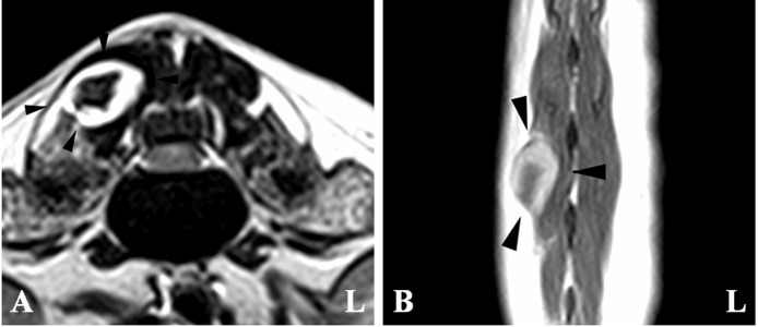
An 11-year-old male Miniature Dachshund was referred for acute neurological deficits in the pelvic limbs. T2-weighted magnetic resonance imaging revealed that the spinal cord at the L1-2 intervertebral disc space was heterogeneously hyperintense in the sagittal plane and was mildly compressed from the ventral side by a small hypointense mass in the transverse plane. However, the lesion showed mass enhancement and severe spinal cord compression on post-contrast T1-weighted imaging. On three-dimensional myelography, a "golf tee sign" was observed around the mass. Therefore, we diagnosed an intradural extramedullary lesion. The mass was surgically removed and histologically diagnosed as a hemangiosarcoma. The "golf tee sign" observed on magnetic resonance myelography may be useful for distinguishing intradural extramedullary masses from intramedullary masses.
一只11岁的雄性迷你腊肠犬因后肢急性神经功能缺损前来就诊。T2加权磁共振成像显示,L1-2椎间盘间隙水平的脊髓在矢状面上呈不均匀高信号,在横断面上被一个小的低信号肿块从腹侧轻度压迫。然而,在增强后T1加权成像上,病变显示肿块强化及严重的脊髓压迫。在三维脊髓造影上,肿块周围观察到“高尔夫球座征”。因此,我们诊断为硬膜内髓外病变。肿块经手术切除,组织学诊断为血管肉瘤。磁共振脊髓造影上观察到的“高尔夫球座征”可能有助于鉴别硬膜内髓外肿块与髓内肿块。