Kang Young-Hoon, Shivakumar Sharath Belame, Son Young-Bum, Bharti Dinesh, Jang Si-Jung, Heo Kang-Sun, Park Won-Uk, Byun June-Ho, Park Bong-Wook, Rho Gyu-Jin
Department of Dentistry, Gyeongsang National University School of Medicine and Institute of Health Science, Jinju, Republic of Korea.
Department of Oral and Maxillofacial Surgery, Changwon Gyeongsang National University Hospital, Changwon, Republic of Korea.
Anim Cells Syst (Seoul). 2019 Jun 10;23(4):275-287. doi: 10.1080/19768354.2019.1626280. eCollection 2019.
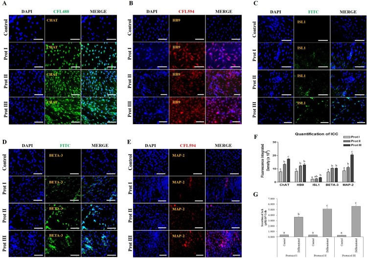
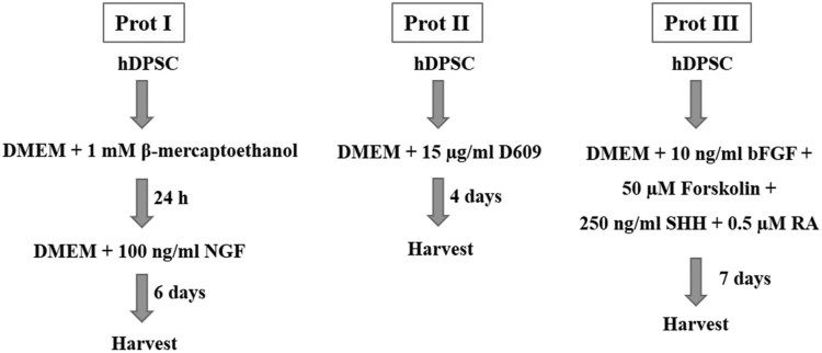
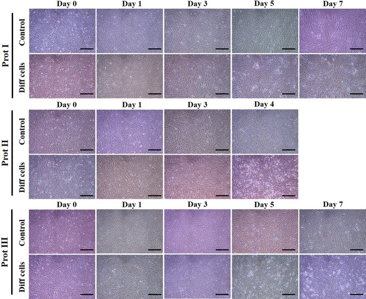
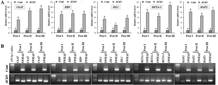
A decrease in the activity of choline acetyltransferase, the enzyme responsible for acetylcholine synthesis in the cholinergic neurons cause neurological disorders involving a decline in cognitive abilities, such as Alzheimer's disease. Mesenchymal stem cells (MSCs) can be used as an efficient therapeutic agents due to their neuronal differentiation potential. Different source derived MSCs may have different differentiation potential under different inductions. Various in vitro protocols have been developed to differentiate MSCs into specific neurons but the comparative effect of different protocols utilizing same source derived MSCs, is not known. To address this issue, dental pulp derived MSCs (DPSCs) were differentiated into cholinergic neurons using three different protocols. In protocol I, DPSCs were pre-induced with serum-free ADMEM containing 1 mM of β-mercaptoethanol for 24 h and then incubated with 100 ng/ml nerve growth factor (NGF) for 6 days. Under protocol II, DPSCs were cultured in serum-free ADMEM containing 15 µg/ml of D609 (tricyclodecan-9-yl-xanthogenate) for 4 days. Under protocol III, the DPSCs were cultured in serum-free ADMEM containing 10 ng/ml of basic fibroblast growth factor (bFGF), 50 µM of forskolin, 250 ng/ml of sonic hedgehog (SHH), and 0.5 µM of retinoic acid (RA) for 7 days. The DPSCs were successfully trans-differentiated under all the protocols, exhibited neuron-like morphologies with upregulated cholinergic neuron-specific markers such as ChAT, HB9, ISL1, BETA-3, and MAP2 both at mRNA and protein levels in comparison to untreated cells. However, protocol III-induced cells showed the highest expression of the cholinergic markers and secreted the highest level of acetylcholine.
胆碱乙酰转移酶是胆碱能神经元中负责合成乙酰胆碱的酶,其活性降低会导致神经功能障碍,包括认知能力下降,如阿尔茨海默病。间充质干细胞(MSCs)由于其神经元分化潜能,可作为一种有效的治疗剂。不同来源的MSCs在不同诱导条件下可能具有不同的分化潜能。已经开发了各种体外方案来将MSCs分化为特定的神经元,但利用相同来源的MSCs的不同方案的比较效果尚不清楚。为了解决这个问题,使用三种不同的方案将牙髓来源的MSCs(DPSCs)分化为胆碱能神经元。在方案I中,DPSCs先用含有1 mMβ-巯基乙醇的无血清ADMEM预诱导24小时,然后与100 ng/ml神经生长因子(NGF)孵育6天。在方案II下,DPSCs在含有15μg/ml D609(三环癸烷-9-基-黄原酸盐)的无血清ADMEM中培养4天。在方案III下,DPSCs在含有10 ng/ml碱性成纤维细胞生长因子(bFGF)、50μM福司可林、250 ng/ml音猬因子(SHH)和0.5μM视黄酸(RA)的无血清ADMEM中培养7天。在所有方案下,DPSCs均成功转分化,与未处理的细胞相比,在mRNA和蛋白质水平上均表现出神经元样形态,胆碱能神经元特异性标志物如ChAT、HB9、ISL1、BETA-3和MAP2上调。然而,方案III诱导的细胞显示胆碱能标志物的表达最高,并且分泌的乙酰胆碱水平最高。