• 文献检索
  • 文档翻译
  • 深度研究
  • 学术资讯
  • Suppr Zotero 插件Zotero 插件
  • 邀请有礼
  • 套餐&价格
  • 历史记录
应用&插件
Suppr Zotero 插件Zotero 插件浏览器插件Mac 客户端Windows 客户端微信小程序
定价
高级版会员购买积分包购买API积分包
服务
文献检索文档翻译深度研究API 文档MCP 服务
关于我们
关于 Suppr公司介绍联系我们用户协议隐私条款
关注我们

Suppr 超能文献

核心技术专利:CN118964589B侵权必究
粤ICP备2023148730 号-1Suppr @ 2026

文献检索

告别复杂PubMed语法,用中文像聊天一样搜索,搜遍4000万医学文献。AI智能推荐,让科研检索更轻松。

立即免费搜索

文件翻译

保留排版,准确专业,支持PDF/Word/PPT等文件格式,支持 12+语言互译。

免费翻译文档

深度研究

AI帮你快速写综述,25分钟生成高质量综述,智能提取关键信息,辅助科研写作。

立即免费体验

基于个体的椎动脉-基底动脉(VA-BA)交界处的血流场的 PIV 研究。

PIV investigation of the flow fields in subject-specific vertebro-basilar (VA-BA) junction.

机构信息

School of Energy and Power Engineering, Xi'an Jiaotong University, No. 28 Xian Ning West Road, Xi'an, 710049, China.

Department of Radiology and Medical Imaging, The First Affiliated Hospital of Xi'an Jiaotong University, 277 Yanta Weest Road, Xi'an, 710061, China.

出版信息

Biomed Eng Online. 2019 Sep 6;18(1):93. doi: 10.1186/s12938-019-0711-9.

DOI:10.1186/s12938-019-0711-9
PMID:31492145
原文链接:https://pmc.ncbi.nlm.nih.gov/articles/PMC6731569/
Abstract

BACKGROUND

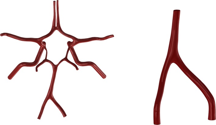
As the only arterial structure of which two main arteries merged into one, the vertebro-basilar (VA-BA) system is one of the favorite sites of cerebral atherosclerotic plaques. The aim of this study was to investigate the detailed hemodynamics characteristics in the VA-BA system.

METHODS

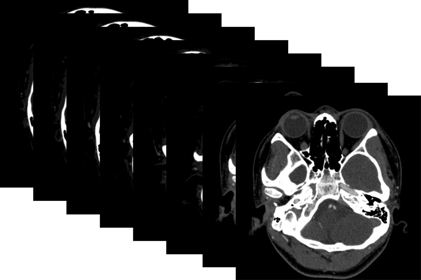
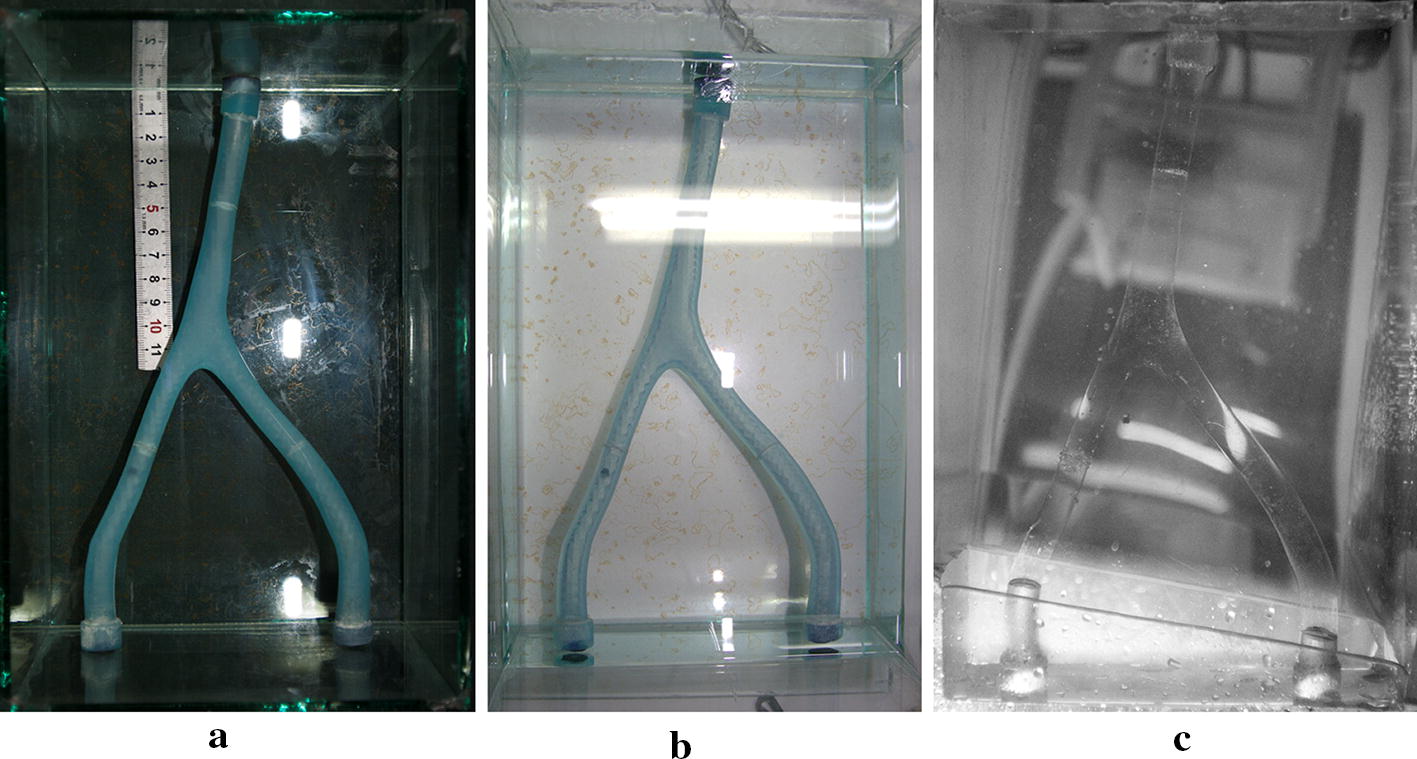
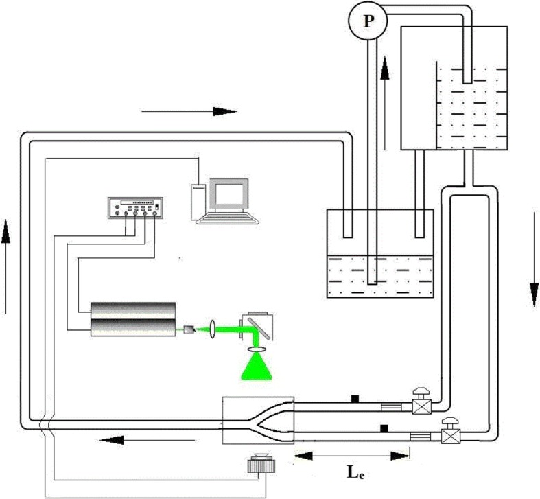
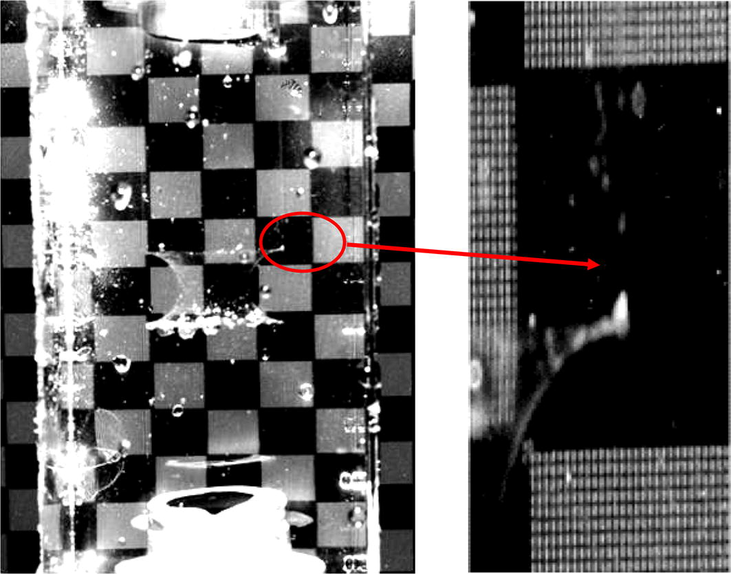
A scale-up subject-specific flow phantom of VA-BA system was fabricated based on the computed tomography angiography (CTA) scanning images of a healthy adult. Flow fields in eight axial planes and six radial planes were measured and analyzed by using particle image velocimetry (PIV) under steady flow conditions of [Formula: see text], [Formula: see text]. A water-glycerin mixture was used as the working fluid.

RESULTS

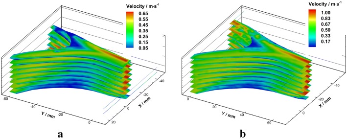
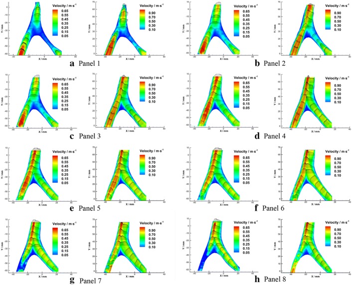
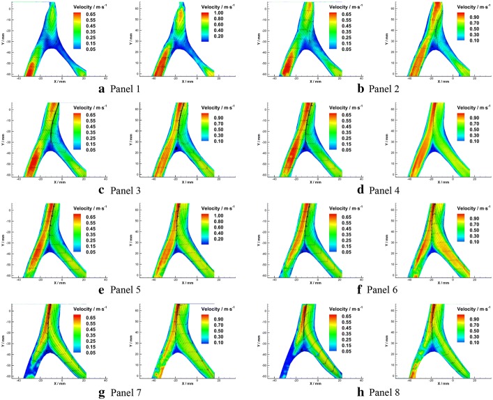
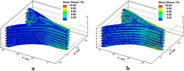
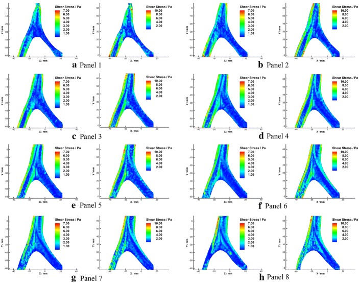
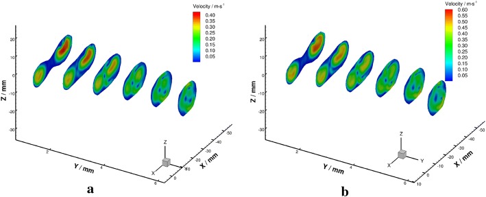
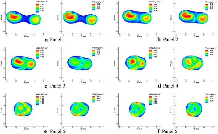
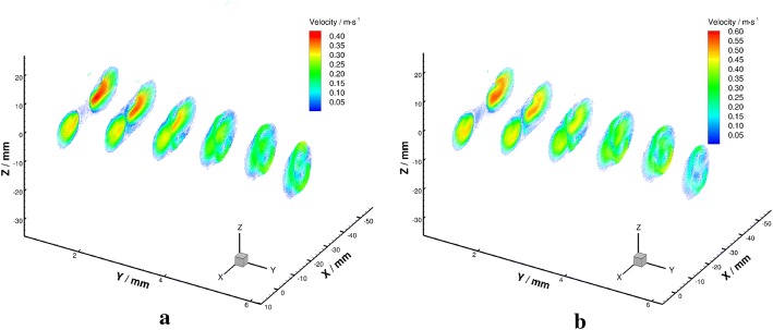
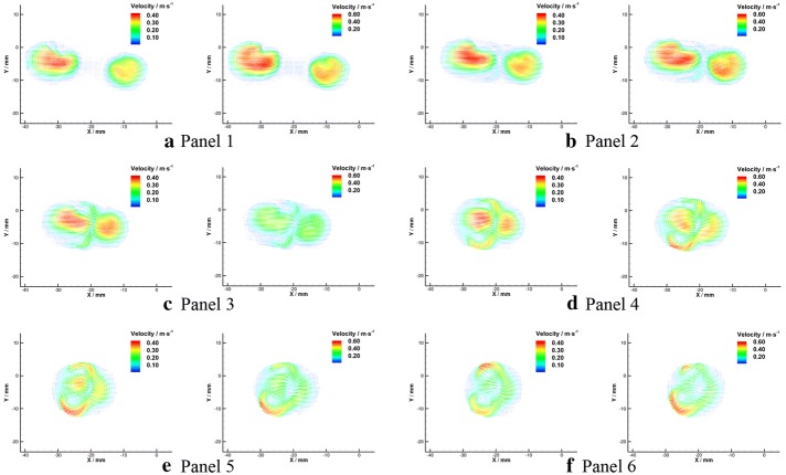
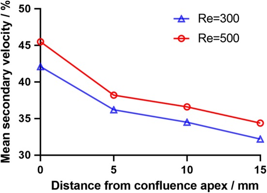
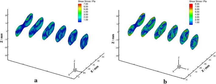
The flow in the current model exhibited highly three-dimensional characteristics. The confluence of VAs flow formed bimodal velocity distribution near the confluence apex. Due to the asymmetrical structural configuration, the bimodal velocity profile skewed towards left, and sharper peaks were observed under higher Reynolds condition. Secondary flow characterized by two vortices formed in the radial planes where 10 mm downstream the confluence apex and persists along the BA under both Reynolds numbers. The strength of secondary flow under [Formula: see text] is around 8% higher than that under [Formula: see text], and decayed nonlinearly along the flow direction. In addition, a low momentum recirculation region induced by boundary layer separation was observed near the confluence apex. The wall shear stress (WSS) in the recirculation area was found to be lower than 0.4 Pa. This region coincides well with the preferential site of vascular lesions in the VA-BA system.

CONCLUSIONS

This preliminary study verified that the subject-specific in-vitro experiment is capable of reflecting the detailed flow features in the VA-BA system. The findings from this study may help to expand the understanding of the hemodynamics in the VA-BA system, and further clarifying the mechanism that underlying the localization of vascular lesions.

摘要

背景

作为两条主要动脉合并为一条的唯一动脉结构,椎基底动脉(VA-BA)系统是脑动脉粥样硬化斑块的最爱部位之一。本研究旨在研究 VA-BA 系统中的详细血液动力学特征。

方法

基于健康成年人的计算机断层血管造影(CTA)扫描图像,制作了一个比例放大的 VA-BA 系统的特定于主体的流量模型。在稳态流条件下,使用粒子图像测速(PIV)测量和分析了八个轴向平面和六个径向平面中的流场,[Formula: see text],[Formula: see text]。水-甘油混合物用作工作流体。

结果

当前模型中的流动表现出高度的三维特征。VA 流的汇流在汇流顶点附近形成了双峰速度分布。由于不对称的结构配置,双峰速度分布向左倾斜,在较高雷诺数条件下观察到更陡峭的峰值。在汇流顶点下游 10mm 的径向平面中形成了以两个涡流为特征的二次流,并在两个雷诺数下沿 BA 持续存在。在[Formula: see text]下的二次流强度比在[Formula: see text]下高约 8%,并沿流动方向呈非线性衰减。此外,在汇流顶点附近观察到边界层分离引起的低动量回流区。发现回流区的壁面剪切应力(WSS)低于 0.4Pa。该区域与 VA-BA 系统中血管病变的优先部位非常吻合。

结论

这项初步研究验证了特定于主体的体外实验能够反映 VA-BA 系统中的详细流动特征。本研究的结果可能有助于扩大对 VA-BA 系统中血液动力学的理解,并进一步阐明血管病变定位的机制。

https://cdn.ncbi.nlm.nih.gov/pmc/blobs/f9af/6731569/4b7c49bc0661/12938_2019_711_Fig18_HTML.jpg
https://cdn.ncbi.nlm.nih.gov/pmc/blobs/f9af/6731569/0fb7a7a12111/12938_2019_711_Fig1_HTML.jpg
https://cdn.ncbi.nlm.nih.gov/pmc/blobs/f9af/6731569/fb1c6972b500/12938_2019_711_Fig2_HTML.jpg
https://cdn.ncbi.nlm.nih.gov/pmc/blobs/f9af/6731569/3f49bad2b43f/12938_2019_711_Fig3_HTML.jpg
https://cdn.ncbi.nlm.nih.gov/pmc/blobs/f9af/6731569/218a9055ffbb/12938_2019_711_Fig4_HTML.jpg
https://cdn.ncbi.nlm.nih.gov/pmc/blobs/f9af/6731569/5561a4939f1c/12938_2019_711_Fig5_HTML.jpg
https://cdn.ncbi.nlm.nih.gov/pmc/blobs/f9af/6731569/a5e7ecb906b9/12938_2019_711_Fig6_HTML.jpg
https://cdn.ncbi.nlm.nih.gov/pmc/blobs/f9af/6731569/1ef286ee229c/12938_2019_711_Fig7_HTML.jpg
https://cdn.ncbi.nlm.nih.gov/pmc/blobs/f9af/6731569/a50c214a4514/12938_2019_711_Fig8_HTML.jpg
https://cdn.ncbi.nlm.nih.gov/pmc/blobs/f9af/6731569/c2948243886d/12938_2019_711_Fig9_HTML.jpg
https://cdn.ncbi.nlm.nih.gov/pmc/blobs/f9af/6731569/6ba14cba2c1b/12938_2019_711_Fig10_HTML.jpg
https://cdn.ncbi.nlm.nih.gov/pmc/blobs/f9af/6731569/20acb0a81b05/12938_2019_711_Fig11_HTML.jpg
https://cdn.ncbi.nlm.nih.gov/pmc/blobs/f9af/6731569/f722ac4fa9db/12938_2019_711_Fig12_HTML.jpg
https://cdn.ncbi.nlm.nih.gov/pmc/blobs/f9af/6731569/88532afbc4e8/12938_2019_711_Fig13_HTML.jpg
https://cdn.ncbi.nlm.nih.gov/pmc/blobs/f9af/6731569/75cb02b8cd04/12938_2019_711_Fig14_HTML.jpg
https://cdn.ncbi.nlm.nih.gov/pmc/blobs/f9af/6731569/bd83002d9681/12938_2019_711_Fig15_HTML.jpg
https://cdn.ncbi.nlm.nih.gov/pmc/blobs/f9af/6731569/3ff8f7856504/12938_2019_711_Fig16_HTML.jpg
https://cdn.ncbi.nlm.nih.gov/pmc/blobs/f9af/6731569/a64c85153fa1/12938_2019_711_Fig17_HTML.jpg
https://cdn.ncbi.nlm.nih.gov/pmc/blobs/f9af/6731569/4b7c49bc0661/12938_2019_711_Fig18_HTML.jpg

相似文献

1
PIV investigation of the flow fields in subject-specific vertebro-basilar (VA-BA) junction.基于个体的椎动脉-基底动脉(VA-BA)交界处的血流场的 PIV 研究。
Biomed Eng Online. 2019 Sep 6;18(1):93. doi: 10.1186/s12938-019-0711-9.
2
Development of an Experimental and Digital Cardiovascular Arterial Model for Transient Hemodynamic and Postural Change Studies: "A Preliminary Framework Analysis".用于瞬态血流动力学和姿势变化研究的实验性和数字化心血管动脉模型的开发:“初步框架分析”
Cardiovasc Eng Technol. 2018 Mar;9(1):1-31. doi: 10.1007/s13239-017-0332-z. Epub 2017 Nov 9.
3
The influence of the angle of confluence on the flow in a vertebro-basilar junction model.汇合角度对椎基底动脉交界区模型中血流的影响。
J Biomech. 1996 Mar;29(3):281-99. doi: 10.1016/0021-9290(95)00064-x.
4
Formation of Vortices in Idealised Branching Vessels: A CFD Benchmark Study.理想化分支血管中涡旋的形成:一项计算流体动力学基准研究。
Cardiovasc Eng Technol. 2020 Oct;11(5):544-559. doi: 10.1007/s13239-020-00477-9. Epub 2020 Jul 14.
5
The influence of blood velocity and vessel geometric parameters on wall shear stress.血流速度和血管几何参数对壁切应力的影响。
Med Eng Phys. 2024 Feb;124:104112. doi: 10.1016/j.medengphy.2024.104112. Epub 2024 Jan 28.
6
PIV-measured versus CFD-predicted flow dynamics in anatomically realistic cerebral aneurysm models.在解剖学逼真的脑动脉瘤模型中,粒子图像测速(PIV)测量与计算流体动力学(CFD)预测的血流动力学对比
J Biomech Eng. 2008 Apr;130(2):021015. doi: 10.1115/1.2900724.
7
Steady Flow in a Patient-Averaged Inferior Vena Cava-Part II: Computational Fluid Dynamics Verification and Validation.患者平均下腔静脉中的稳定血流 - 第二部分:计算流体动力学验证与确认
Cardiovasc Eng Technol. 2018 Dec;9(4):654-673. doi: 10.1007/s13239-018-00392-0. Epub 2018 Nov 16.
8
Computational and experimental assessment of influences of hemodynamic shear stress on carotid plaque.血流动力学剪切应力对颈动脉斑块影响的计算与实验评估
Biomed Eng Online. 2017 Jul 29;16(1):92. doi: 10.1186/s12938-017-0386-z.
9
Towards the prediction of flow-induced shear stress distributions experienced by breast cancer cells in the lymphatics.旨在预测乳腺癌细胞在淋巴管中所经历的流致切应力分布。
Biomech Model Mechanobiol. 2017 Dec;16(6):2051-2062. doi: 10.1007/s10237-017-0937-z. Epub 2017 Jul 24.
10
Measurement of Wall Shear Stress Exerted by Flowing Blood in the Human Carotid Artery: Ultrasound Doppler Velocimetry and Echo Particle Image Velocimetry.人体颈动脉中流动血液所施加的壁面剪应力测量:超声多普勒测速法与回声粒子图像测速法。
Ultrasound Med Biol. 2018 Jul;44(7):1392-1401. doi: 10.1016/j.ultrasmedbio.2018.02.013. Epub 2018 Apr 17.

本文引用的文献

1
Relationship between the geometry patterns of vertebrobasilar artery and atherosclerosis.椎基底动脉几何形态与动脉粥样硬化之间的关系。
BMC Neurol. 2018 Jun 12;18(1):83. doi: 10.1186/s12883-018-1084-6.
2
Three-dimensional flow structures past a bio-prosthetic valve in an in-vitro model of the aortic root.体外主动脉窦模型中生物假体瓣周围的三维流结构。
PLoS One. 2018 Mar 16;13(3):e0194384. doi: 10.1371/journal.pone.0194384. eCollection 2018.
3
Impact of spatial characteristics in the left stenotic coronary artery on the hemodynamics and visualization of 3D replica models.
左冠状动脉狭窄处空间特征对血流动力学及三维复制模型可视化的影响
Sci Rep. 2017 Nov 13;7(1):15452. doi: 10.1038/s41598-017-15620-1.
4
Impact of bifurcation angle and other anatomical characteristics on blood flow - A computational study of non-stented and stented coronary arteries.分叉角度及其他解剖学特征对血流的影响——无支架和有支架冠状动脉的计算研究
J Biomech. 2016 Jun 14;49(9):1570-1582. doi: 10.1016/j.jbiomech.2016.03.038. Epub 2016 Mar 29.
5
Three-dimensional hemodynamics analysis of the circle of Willis in the patient-specific nonintegral arterial structures.特定患者非整体动脉结构中 Willis 环的三维血流动力学分析。
Biomech Model Mechanobiol. 2016 Dec;15(6):1439-1456. doi: 10.1007/s10237-016-0773-6. Epub 2016 Mar 3.
6
The role of the circle of Willis in internal carotid artery stenosis and anatomical variations: a computational study based on a patient-specific three-dimensional model.Willis环在颈内动脉狭窄及解剖变异中的作用:基于个体化三维模型的计算研究
Biomed Eng Online. 2015 Nov 25;14:107. doi: 10.1186/s12938-015-0105-6.
7
Hemodynamics in growing and stable cerebral aneurysms.生长中和稳定期脑动脉瘤的血流动力学
J Neurointerv Surg. 2016 Apr;8(4):407-12. doi: 10.1136/neurintsurg-2014-011339. Epub 2015 Feb 4.
8
Experimental study of hemodynamics in the Circle of Willis.Willis 环血流动力学的实验研究
Biomed Eng Online. 2015;14 Suppl 1(Suppl 1):S10. doi: 10.1186/1475-925X-14-S1-S10. Epub 2015 Jan 9.
9
Vertebro-basilar junction aneurysms: a single centre experience and meta-analysis of endovascular treatments.椎基底动脉交界处动脉瘤:单中心经验及血管内治疗的荟萃分析
Neuroradiol J. 2014 Dec;27(6):732-41. doi: 10.15274/NRJ-2014-10100. Epub 2014 Dec 1.
10
Change of direction in the biomechanics of atherosclerosis.动脉粥样硬化生物力学中的方向转变。
Ann Biomed Eng. 2015 Jan;43(1):16-25. doi: 10.1007/s10439-014-1095-4. Epub 2014 Aug 20.