Li Jiahui, Liu Huimin, Mauer Amy S, Lucien Fabrice, Raiter Abagail, Bandla Harikrishna, Mounajjed Taofic, Yin Ziying, Glaser Kevin J, Yin Meng, Malhi Harmeet
Department of Radiology Mayo Clinic Rochester MN.
Division of Gastroenterology and Hepatology Mayo Clinic Rochester MN.
Hepatol Commun. 2019 Jul 10;3(9):1235-1249. doi: 10.1002/hep4.1404. eCollection 2019 Sep.
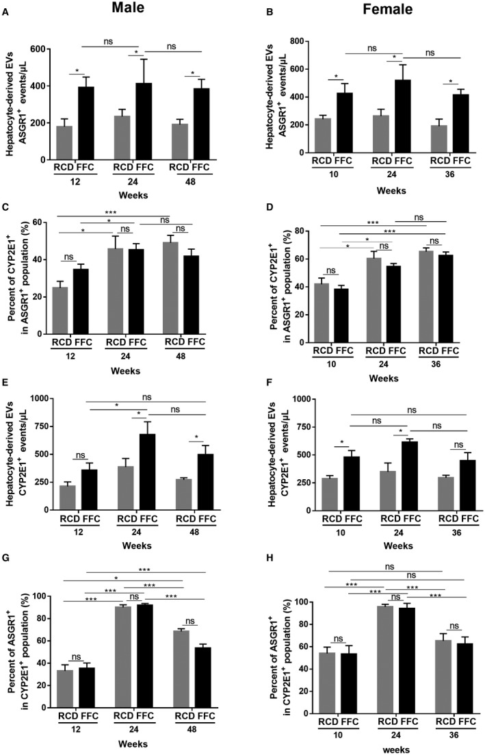
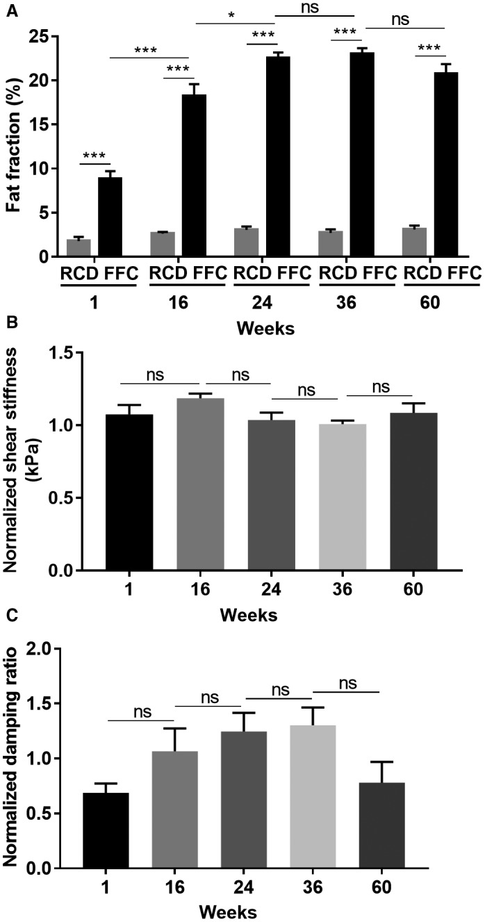
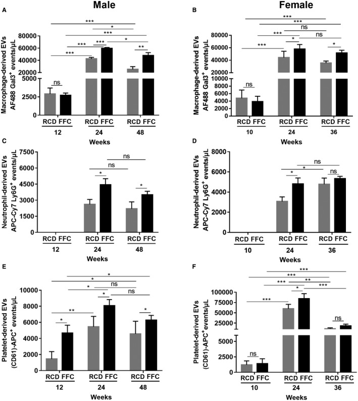
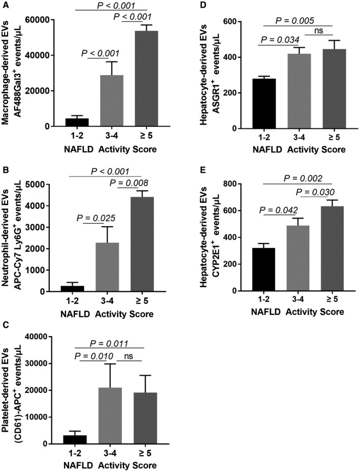
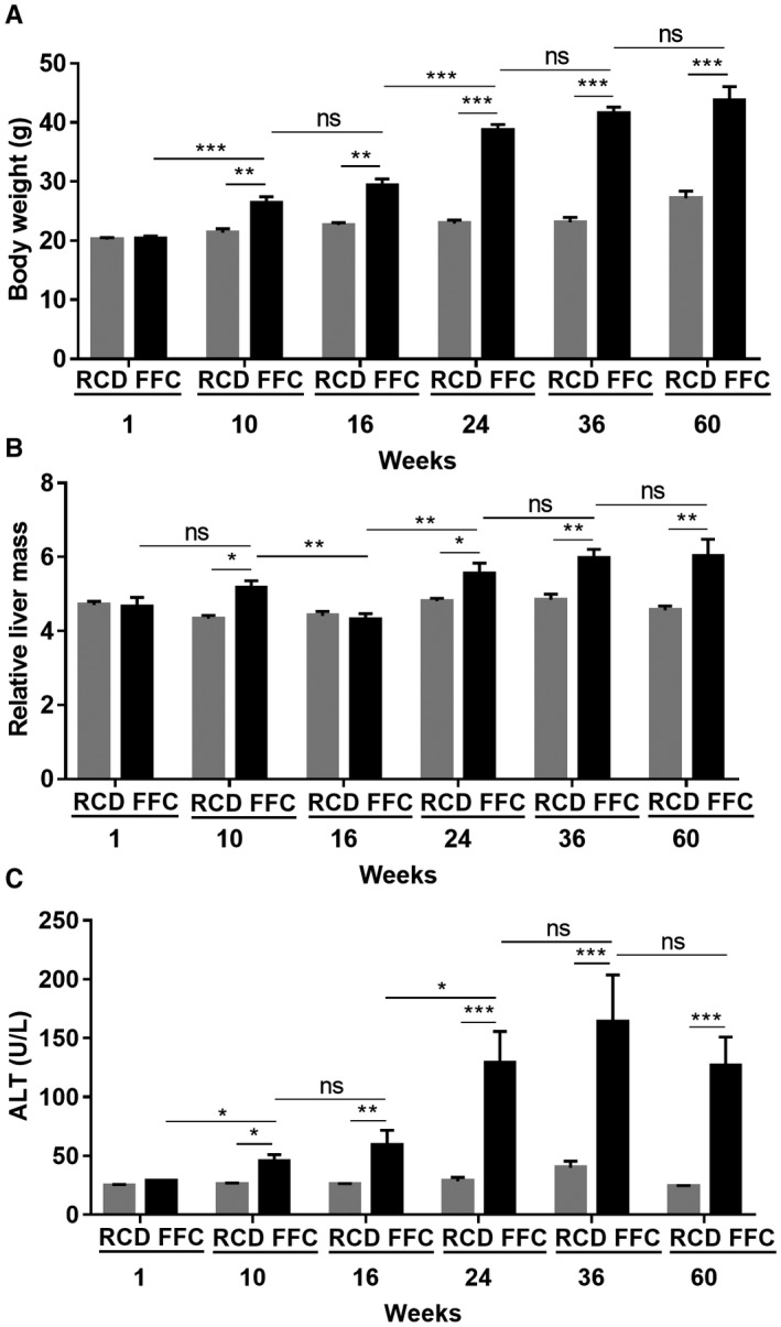
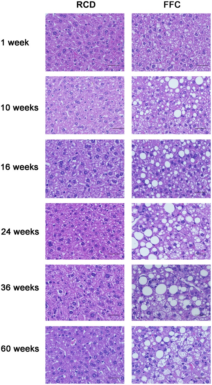
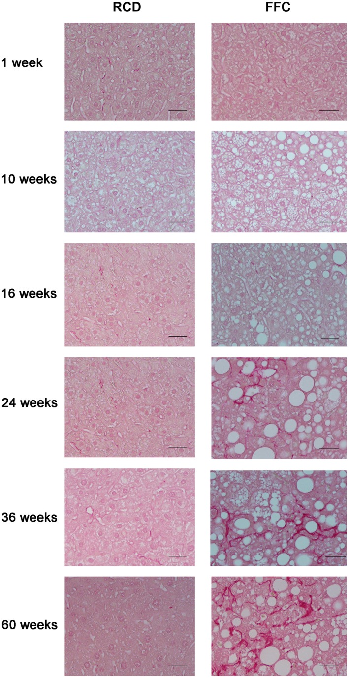
Circulating extracellular vesicles (EVs) are a novel and emerging biomarker for nonalcoholic steatohepatitis (NASH). It has been demonstrated that total circulating EVs and hepatocyte-derived EVs are elevated in male mice with diet-induced NASH. How hepatocyte-derived EVs change over time and other cellular sources of EVs in NASH have not been determined. Our objective was to define the quantitative evolution of hepatocyte-derived, macrophage-derived, neutrophil-derived, and platelet-derived EVs in male and female mice with dietary NASH. Fluorescently labeled antibodies and a nanoscale flow cytometer were used to detect plasma levels of EVs. Asialoglycoprotein receptor 1 (ASGR1) and cytochrome P450 family 2 subfamily E member 1 (CYP2E1) are markers of hepatocyte-derived EVs; galectin 3 is a marker of macrophage-derived EVs; common epitope on lymphocyte antigen 6 complex, locus G/C1 (Ly-6G and Ly-6C) is a marker of neutrophil-derived EVs; and clusters of differentiation 61 (CD61) is a marker of platelet-derived EVs. Nonalcoholic fatty liver disease activity score (NAS) was calculated using hematoxylin and eosin-stained liver sections, and magnetic resonance imaging (MRI) was used for measurement of the fat fraction and elastography. Hepatocyte-derived EVs increased in both male and female mice at 12 and 10 weeks of feeding, respectively, and remained elevated at 24 weeks in both male and female mice and at 48 weeks in male mice and 36 weeks in female mice. Macrophage- and neutrophil-derived EVs were significantly elevated at 24 weeks of dietary feeding concomitant with the histologic presence of inflammatory foci in the liver. In fat-, fructose-, and cholesterol- (FFC) fed male mice, platelet-derived EVs were elevated at 12, 24, and 48 weeks, whereas in female mice, platelet derived EVs were significantly elevated at 24 weeks. Hepatocyte-, macrophage- and neutrophil-derived EVs correlated well with the histologic NAS. Circulating cell-type-specific EVs may be a novel biomarker for NASH diagnosis and longitudinal follow up.
循环细胞外囊泡(EVs)是一种用于非酒精性脂肪性肝炎(NASH)的新型且正在兴起的生物标志物。已证实,在饮食诱导的NASH雄性小鼠中,循环总EVs和肝细胞来源的EVs升高。肝细胞来源的EVs如何随时间变化以及NASH中EVs的其他细胞来源尚未确定。我们的目标是确定饮食诱导的NASH雄性和雌性小鼠中肝细胞来源、巨噬细胞来源、中性粒细胞来源和血小板来源的EVs的定量演变。使用荧光标记抗体和纳米级流式细胞仪检测EVs的血浆水平。去唾液酸糖蛋白受体1(ASGR1)和细胞色素P450家族2亚家族E成员1(CYP2E1)是肝细胞来源的EVs的标志物;半乳糖凝集素3是巨噬细胞来源的EVs的标志物;淋巴细胞抗原6复合体G/C1位点(Ly-6G和Ly-6C)上的共同表位是中性粒细胞来源的EVs的标志物;分化簇61(CD61)是血小板来源的EVs的标志物。使用苏木精和伊红染色的肝脏切片计算非酒精性脂肪性肝病活动评分(NAS),并使用磁共振成像(MRI)测量脂肪分数和弹性成像。肝细胞来源的EVs在雄性和雌性小鼠分别喂养12周和10周时增加,并在雄性和雌性小鼠24周时、雄性小鼠48周时和雌性小鼠36周时保持升高。巨噬细胞和中性粒细胞来源的EVs在饮食喂养24周时显著升高,并伴有肝脏中炎症灶的组织学存在。在喂食脂肪、果糖和胆固醇(FFC)的雄性小鼠中,血小板来源的EVs在12周、24周和48周时升高,而在雌性小鼠中,血小板来源的EVs在24周时显著升高。肝细胞、巨噬细胞和中性粒细胞来源的EVs与组织学NAS密切相关。循环细胞类型特异性EVs可能是NASH诊断和纵向随访的新型生物标志物。