Department of Oncology, The First Affiliated Hospital of Nanjing Medical University, Nanjing 210029, China.
State Key Laboratory of Pharmaceutical Biotechnology and Collaborative Innovation Center of Chemistry for Life Sciences, School of Life Sciences, Nanjing University, Nanjing 210023, China.
EBioMedicine. 2019 Oct;48:169-177. doi: 10.1016/j.ebiom.2019.08.067. Epub 2019 Sep 11.
Tumor mutations and tumor microenvironment are associated with resistance to cancer immunotherapies. However, peripheral T cell in effective anti-programmed death 1 (PD-1) antibody treatment is poorly understood.
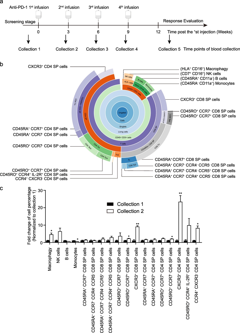
Mass spectrometry and conventional flow cytometry were used to investigate peripheral blood cells isolated from patients. Furthermore, melanoma mouse model was performed to assess the role of CXCR3 signaling in anti-PD-1 antibody treatment.
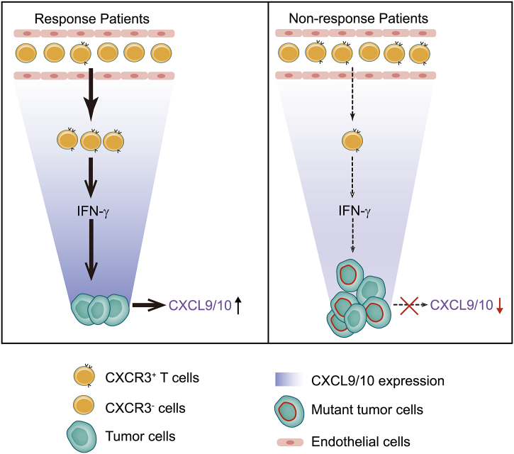
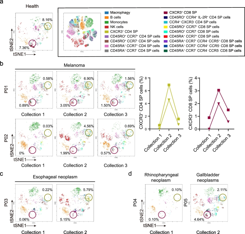
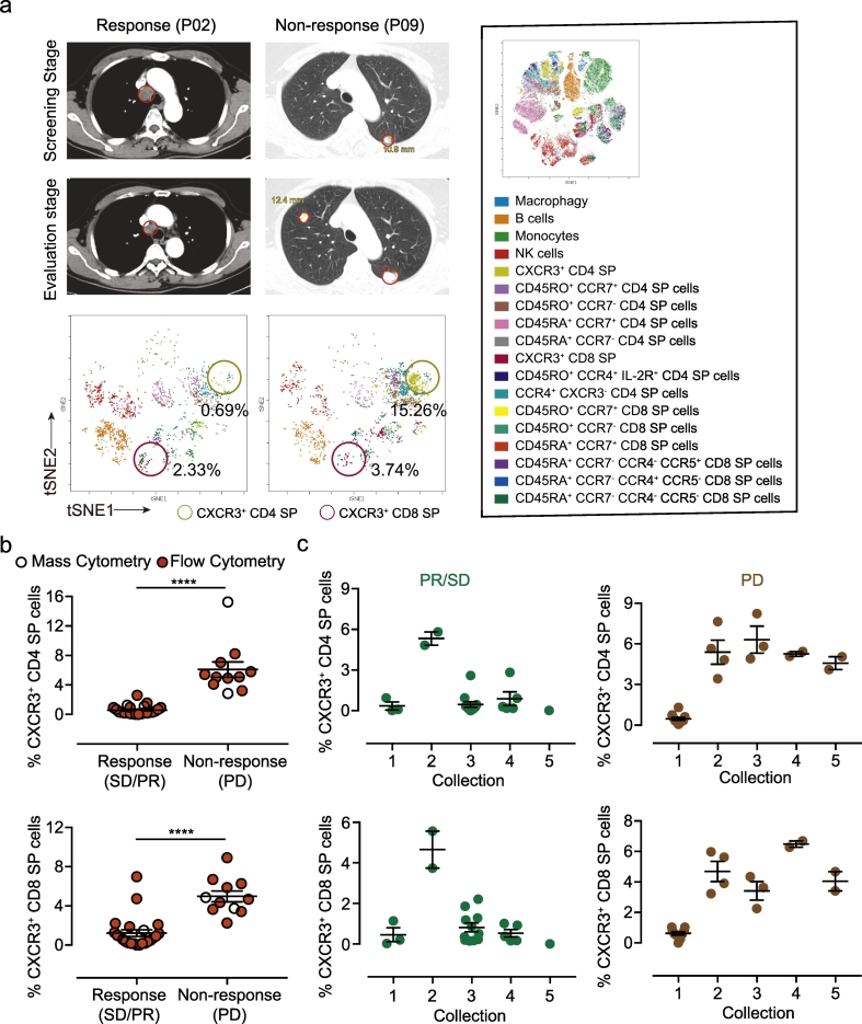
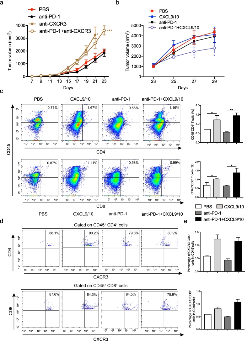
We revealed a marked increase in the percentage of CXCR3 T cells in the blood of cancer patients after the first pembrolizumab infusion. This percentage decreased after the second infusion in responsive patients, whereas a sustained high percentage of CXCR3 T cells was observed in patients with progressive disease. A low percentage of CXCR3 T cells presented in patients with stable disease or a partial response was confirmed by conventional flow cytometry. Intriguingly, blockade of CXCR3 signaling exacerbated tumor growth in mice. Intratumoral injection with recombinant CXCL9/10 plus intraperitoneal injection of anti-PD1 antibody inhibited the tumor growth in mice.
The dynamic changes in CXCR3 T cells in blood may be a prognostic factor in anti-PD-1 immunotherapy, and promotion of CXCR3-mediated signaling may be beneficial to the anti-PD-1 therapy. FUND: This work was supported by the National Natural Science Foundation of China (Nos. 81722047, 81871944, 81670553, 81874317, 81572389, 81730100) and Jiangsu province key medical talents (Nos. ZDRCA2016026), The "Deng Feng" Distinguished Scholars Program, National Science & Technology Major Project "Key New Drug Creation and Manufacturing Program", China (Number: 2018ZX09201002), and the Fundamental Research Funds for the Central Universities (020814380117).
肿瘤突变和肿瘤微环境与癌症免疫疗法的耐药性有关。然而,外周 T 细胞在有效的抗程序性死亡 1(PD-1)抗体治疗中的作用仍不清楚。
使用质谱和常规流式细胞术来研究从患者中分离的外周血细胞。此外,进行黑色素瘤小鼠模型以评估 CXCR3 信号在抗 PD-1 抗体治疗中的作用。
我们发现,在接受帕博利珠单抗首次输注后,癌症患者血液中 CXCR3 T 细胞的百分比明显增加。在有反应的患者中,第二次输注后该百分比降低,而在疾病进展的患者中则观察到 CXCR3 T 细胞的持续高百分比。常规流式细胞术证实,在疾病稳定或部分缓解的患者中,CXCR3 T 细胞的百分比较低。有趣的是,阻断 CXCR3 信号会加剧小鼠的肿瘤生长。肿瘤内注射重组 CXCL9/10 加腹腔注射抗 PD-1 抗体抑制了小鼠的肿瘤生长。
血液中 CXCR3 T 细胞的动态变化可能是抗 PD-1 免疫治疗的预后因素,促进 CXCR3 介导的信号可能有利于抗 PD-1 治疗。
本工作得到了国家自然科学基金(Nos. 81722047、81871944、81670553、81730100、81572389、81770553)、江苏省重点医学人才(Nos. ZDRCA2016026)、“邓峰”杰出学者计划、国家科技重大专项“重大新药创制”(编号:2018ZX09201002)和中央高校基本科研业务费(020814380117)的支持。