• 文献检索
  • 文档翻译
  • 深度研究
  • 学术资讯
  • Suppr Zotero 插件Zotero 插件
  • 邀请有礼
  • 套餐&价格
  • 历史记录
应用&插件
Suppr Zotero 插件Zotero 插件浏览器插件Mac 客户端Windows 客户端微信小程序
定价
高级版会员购买积分包购买API积分包
服务
文献检索文档翻译深度研究API 文档MCP 服务
关于我们
关于 Suppr公司介绍联系我们用户协议隐私条款
关注我们

Suppr 超能文献

核心技术专利:CN118964589B侵权必究
粤ICP备2023148730 号-1Suppr @ 2026

文献检索

告别复杂PubMed语法,用中文像聊天一样搜索,搜遍4000万医学文献。AI智能推荐,让科研检索更轻松。

立即免费搜索

文件翻译

保留排版,准确专业,支持PDF/Word/PPT等文件格式,支持 12+语言互译。

免费翻译文档

深度研究

AI帮你快速写综述,25分钟生成高质量综述,智能提取关键信息,辅助科研写作。

立即免费体验

纯化的 F-ATP 合酶形成了一种依赖 Ca2+的高电导通道,与线粒体通透性转换孔相匹配。

Purified F-ATP synthase forms a Ca-dependent high-conductance channel matching the mitochondrial permeability transition pore.

机构信息

Department of Biomedical Sciences, University of Padova, 35131, Padova, Italy.

Consiglio Nazionale delle Ricerche Neuroscience Institute, 35131, Padova, Italy.

出版信息

Nat Commun. 2019 Sep 25;10(1):4341. doi: 10.1038/s41467-019-12331-1.

DOI:10.1038/s41467-019-12331-1
PMID:31554800
原文链接:https://pmc.ncbi.nlm.nih.gov/articles/PMC6761146/
Abstract

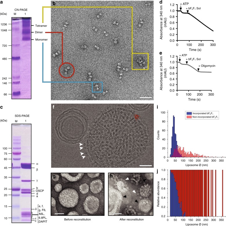
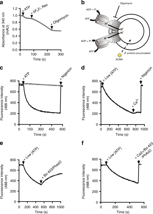
The molecular identity of the mitochondrial megachannel (MMC)/permeability transition pore (PTP), a key effector of cell death, remains controversial. By combining highly purified, fully active bovine F-ATP synthase with preformed liposomes we show that Ca dissipates the H gradient generated by ATP hydrolysis. After incorporation of the same preparation into planar lipid bilayers Ca elicits currents matching those of the MMC/PTP. Currents were fully reversible, were stabilized by benzodiazepine 423, a ligand of the OSCP subunit of F-ATP synthase that activates the MMC/PTP, and were inhibited by Mg and adenine nucleotides, which also inhibit the PTP. Channel activity was insensitive to inhibitors of the adenine nucleotide translocase (ANT) and of the voltage-dependent anion channel (VDAC). Native gel-purified oligomers and dimers, but not monomers, gave rise to channel activity. These findings resolve the long-standing mystery of the MMC/PTP and demonstrate that Ca can transform the energy-conserving F-ATP synthase into an energy-dissipating device.

摘要

线粒体巨大通道(MMC)/通透性转换孔(PTP)是细胞死亡的关键效应因子,其分子身份一直存在争议。通过将高度纯化、完全活性的牛 F-ATP 合酶与预形成的脂质体结合,我们发现 Ca2+ 会耗散由 ATP 水解产生的 H+ 梯度。将相同的制剂掺入平面脂质双层后,Ca2+ 会引发与 MMC/PTP 相匹配的电流。电流是完全可逆的,被 F-ATP 合酶 OSCP 亚基配体苯二氮䓬 423 稳定,该配体能激活 MMC/PTP,并且被 Mg 和腺嘌呤核苷酸抑制,而腺嘌呤核苷酸也能抑制 PTP。通道活性对腺嘌呤核苷酸转运蛋白(ANT)和电压依赖性阴离子通道(VDAC)的抑制剂不敏感。天然凝胶纯化的寡聚体和二聚体,但不是单体,产生了通道活性。这些发现解决了 MMC/PTP 长期存在的谜团,并表明 Ca2+ 可以将能量守恒的 F-ATP 合酶转化为能量耗散装置。

https://cdn.ncbi.nlm.nih.gov/pmc/blobs/d248/6761146/651dcb506db7/41467_2019_12331_Fig5_HTML.jpg
https://cdn.ncbi.nlm.nih.gov/pmc/blobs/d248/6761146/b91a7dae834d/41467_2019_12331_Fig1_HTML.jpg
https://cdn.ncbi.nlm.nih.gov/pmc/blobs/d248/6761146/c0686d25a5cb/41467_2019_12331_Fig2_HTML.jpg
https://cdn.ncbi.nlm.nih.gov/pmc/blobs/d248/6761146/0ab92ffe3056/41467_2019_12331_Fig3_HTML.jpg
https://cdn.ncbi.nlm.nih.gov/pmc/blobs/d248/6761146/5f7e9f0d3222/41467_2019_12331_Fig4_HTML.jpg
https://cdn.ncbi.nlm.nih.gov/pmc/blobs/d248/6761146/651dcb506db7/41467_2019_12331_Fig5_HTML.jpg

相似文献

1
Purified F-ATP synthase forms a Ca-dependent high-conductance channel matching the mitochondrial permeability transition pore.纯化的 F-ATP 合酶形成了一种依赖 Ca2+的高电导通道,与线粒体通透性转换孔相匹配。
Nat Commun. 2019 Sep 25;10(1):4341. doi: 10.1038/s41467-019-12331-1.
2
Ca binding to F-ATP synthase β subunit triggers the mitochondrial permeability transition.钙与F-ATP合酶β亚基结合会引发线粒体通透性转变。
EMBO Rep. 2017 Jul;18(7):1065-1076. doi: 10.15252/embr.201643354. Epub 2017 May 15.
3
Dimers of mitochondrial ATP synthase form the permeability transition pore.线粒体 ATP 合酶二聚体形成了通透性转换孔。
Proc Natl Acad Sci U S A. 2013 Apr 9;110(15):5887-92. doi: 10.1073/pnas.1217823110. Epub 2013 Mar 25.
4
The unique histidine in OSCP subunit of F-ATP synthase mediates inhibition of the permeability transition pore by acidic pH.F-ATP 合酶 OSCP 亚基中的独特组氨酸通过酸性 pH 介导对渗透转换孔的抑制。
EMBO Rep. 2018 Feb;19(2):257-268. doi: 10.15252/embr.201744705. Epub 2017 Dec 7.
5
High-Conductance Channel Formation in Yeast Mitochondria is Mediated by F-ATP Synthase e and g Subunits.酵母线粒体中高电导通道的形成由F-ATP合酶的e和g亚基介导。
Cell Physiol Biochem. 2018;50(5):1840-1855. doi: 10.1159/000494864. Epub 2018 Nov 13.
6
Arg-8 of yeast subunit e contributes to the stability of F-ATP synthase dimers and to the generation of the full-conductance mitochondrial megachannel.酵母亚基 e 的 Arg-8 有助于 F-ATP 合酶二聚体的稳定性和全电导线粒体巨型通道的产生。
J Biol Chem. 2019 Jul 12;294(28):10987-10997. doi: 10.1074/jbc.RA119.008775. Epub 2019 Jun 3.
7
From ATP to PTP and Back: A Dual Function for the Mitochondrial ATP Synthase.从ATP到PTP再返回:线粒体ATP合酶的双重功能
Circ Res. 2015 May 22;116(11):1850-62. doi: 10.1161/CIRCRESAHA.115.306557.
8
F-ATP synthase and the permeability transition pore: fewer doubts, more certainties.F-ATP 合酶与通透性转换孔:更少的疑问,更多的确定。
FEBS Lett. 2019 Jul;593(13):1542-1553. doi: 10.1002/1873-3468.13485. Epub 2019 Jun 24.
9
The mitochondrial permeability transition: Recent progress and open questions.线粒体通透性转换:最新进展与未解问题
FEBS J. 2022 Nov;289(22):7051-7074. doi: 10.1111/febs.16254. Epub 2021 Nov 12.
10
Mitochondrial F-ATP Synthase Co-Migrating Proteins and Ca-Dependent Formation of Large Channels.线粒体F-ATP合酶共迁移蛋白与大通道的钙依赖性形成
Cells. 2023 Oct 7;12(19):2414. doi: 10.3390/cells12192414.

引用本文的文献

1
Cyclophilin D (PPIF) and MPTP in hepatic ischemia-reperfusion injury: insights into mechanisms.亲环素D(PPIF)与肝缺血再灌注损伤中的线粒体通透性转换孔:作用机制剖析
Front Immunol. 2025 Aug 29;16:1575242. doi: 10.3389/fimmu.2025.1575242. eCollection 2025.
2
Comprehensive analysis of regulated cell death pathways: intrinsic disorder, protein-protein interactions, and cross-pathway communication.细胞程序性死亡途径的综合分析:内在无序、蛋白质-蛋白质相互作用及跨途径通讯
Apoptosis. 2025 Aug 19. doi: 10.1007/s10495-025-02161-6.
3
Design and synthesis of 1,4,8-triazaspiro[4.5]decan-2-one derivatives as novel mitochondrial permeability transition pore inhibitors.

本文引用的文献

1
Persistence of the permeability transition pore in human mitochondria devoid of an assembled ATP synthase.人线粒体中缺乏组装的 ATP 合酶时通透性转换孔的持续存在。
Proc Natl Acad Sci U S A. 2019 Jun 25;116(26):12816-12821. doi: 10.1073/pnas.1904005116. Epub 2019 Jun 18.
2
F-ATP synthase and the permeability transition pore: fewer doubts, more certainties.F-ATP 合酶与通透性转换孔:更少的疑问,更多的确定。
FEBS Lett. 2019 Jul;593(13):1542-1553. doi: 10.1002/1873-3468.13485. Epub 2019 Jun 24.
3
Cryo-EM structure of the mammalian ATP synthase tetramer bound with inhibitory protein IF1.
新型线粒体通透性转换孔抑制剂1,4,8-三氮杂螺[4.5]癸-2-酮衍生物的设计与合成
J Enzyme Inhib Med Chem. 2025 Dec;40(1):2505907. doi: 10.1080/14756366.2025.2505907. Epub 2025 May 21.
4
Cryo-EM structure of the brine shrimp mitochondrial ATP synthase suggests an inactivation mechanism for the ATP synthase leak channel.卤虫线粒体ATP合酶的冷冻电镜结构揭示了ATP合酶泄漏通道的失活机制。
Cell Death Differ. 2025 Mar 19. doi: 10.1038/s41418-025-01476-w.
5
N-terminal cleavage of cyclophilin D boosts its ability to bind F-ATP synthase.亲环素 D 的 N 端切割增强了其与 F-ATP 合酶结合的能力。
Commun Biol. 2024 Nov 11;7(1):1486. doi: 10.1038/s42003-024-07172-8.
6
Modulation of mitochondrial permeability transition pores in reperfusion injury: Mechanisms and therapeutic approaches.再灌注损伤中线粒体通透性转换孔的调节:机制与治疗方法。
Eur J Clin Invest. 2025 Jan;55(1):e14331. doi: 10.1111/eci.14331. Epub 2024 Oct 10.
7
Mechanism and treatment of intracerebral hemorrhage focus on mitochondrial permeability transition pore.脑出血的机制与治疗聚焦于线粒体通透性转换孔。
Front Mol Neurosci. 2024 Jul 31;17:1423132. doi: 10.3389/fnmol.2024.1423132. eCollection 2024.
8
Sphingosine is involved in PAPTP-induced death of pancreas cancer cells by interfering with mitochondrial functions.鞘氨醇通过干扰线粒体功能参与 PAPTP 诱导的胰腺癌细胞死亡。
J Mol Med (Berl). 2024 Jul;102(7):947-959. doi: 10.1007/s00109-024-02456-2. Epub 2024 May 23.
9
Pharmaceutical Manipulation of Mitochondrial F0F1-ATP Synthase Enables Imaging and Protection of Myocardial Ischemia/Reperfusion Injury Through Stress-induced Selective Enrichment.药物操控线粒体 F0F1-ATP 合酶通过应激诱导的选择性富集实现心肌缺血/再灌注损伤的成像和保护。
Adv Sci (Weinh). 2024 Mar;11(9):e2307880. doi: 10.1002/advs.202307880. Epub 2023 Dec 14.
10
The mitochondrial ATP synthase is a negative regulator of the mitochondrial permeability transition pore.线粒体 ATP 合酶是线粒体通透性转换孔的负调节剂。
Proc Natl Acad Sci U S A. 2023 Dec 19;120(51):e2303713120. doi: 10.1073/pnas.2303713120. Epub 2023 Dec 13.
哺乳动物 ATP 合酶四聚体与抑制蛋白 IF1 结合的冷冻电镜结构。
Science. 2019 Jun 14;364(6445):1068-1075. doi: 10.1126/science.aaw4852.
4
Arg-8 of yeast subunit e contributes to the stability of F-ATP synthase dimers and to the generation of the full-conductance mitochondrial megachannel.酵母亚基 e 的 Arg-8 有助于 F-ATP 合酶二聚体的稳定性和全电导线粒体巨型通道的产生。
J Biol Chem. 2019 Jul 12;294(28):10987-10997. doi: 10.1074/jbc.RA119.008775. Epub 2019 Jun 3.
5
Cryo-EM reveals distinct conformations of ATP synthase on exposure to ATP.冷冻电镜揭示了在接触 ATP 时 ATP 合酶的不同构象。
Elife. 2019 Mar 26;8:e43864. doi: 10.7554/eLife.43864.
6
Dimers of mitochondrial ATP synthase induce membrane curvature and self-assemble into rows.线粒体 ATP 合酶二聚体诱导膜弯曲,并自组装成排。
Proc Natl Acad Sci U S A. 2019 Mar 5;116(10):4250-4255. doi: 10.1073/pnas.1816556116. Epub 2019 Feb 13.
7
ATP Synthase C-Subunit-Deficient Mitochondria Have a Small Cyclosporine A-Sensitive Channel, but Lack the Permeability Transition Pore.ATP 合酶 C 亚基缺陷型线粒体具有小的环孢素 A 敏感通道,但缺乏通透性转换孔。
Cell Rep. 2019 Jan 2;26(1):11-17.e2. doi: 10.1016/j.celrep.2018.12.033.
8
Properties of the Permeability Transition of Pea Stem Mitochondria.豌豆茎线粒体通透性转换的特性
Front Physiol. 2018 Nov 21;9:1626. doi: 10.3389/fphys.2018.01626. eCollection 2018.
9
Why F-ATP Synthase Remains a Strong Candidate as the Mitochondrial Permeability Transition Pore.为何F-ATP合酶仍是线粒体通透性转换孔的有力候选者。
Front Physiol. 2018 Nov 1;9:1543. doi: 10.3389/fphys.2018.01543. eCollection 2018.
10
High-Conductance Channel Formation in Yeast Mitochondria is Mediated by F-ATP Synthase e and g Subunits.酵母线粒体中高电导通道的形成由F-ATP合酶的e和g亚基介导。
Cell Physiol Biochem. 2018;50(5):1840-1855. doi: 10.1159/000494864. Epub 2018 Nov 13.