Department of Neurosurgery, Oregon Health & Science University, Portland, Oregon, United States of America.
Department of Anesthesiology and Perioperative Medicine, Oregon Health & Science University, Portland, Oregon, United States of America.
PLoS One. 2019 Oct 9;14(10):e0215789. doi: 10.1371/journal.pone.0215789. eCollection 2019.
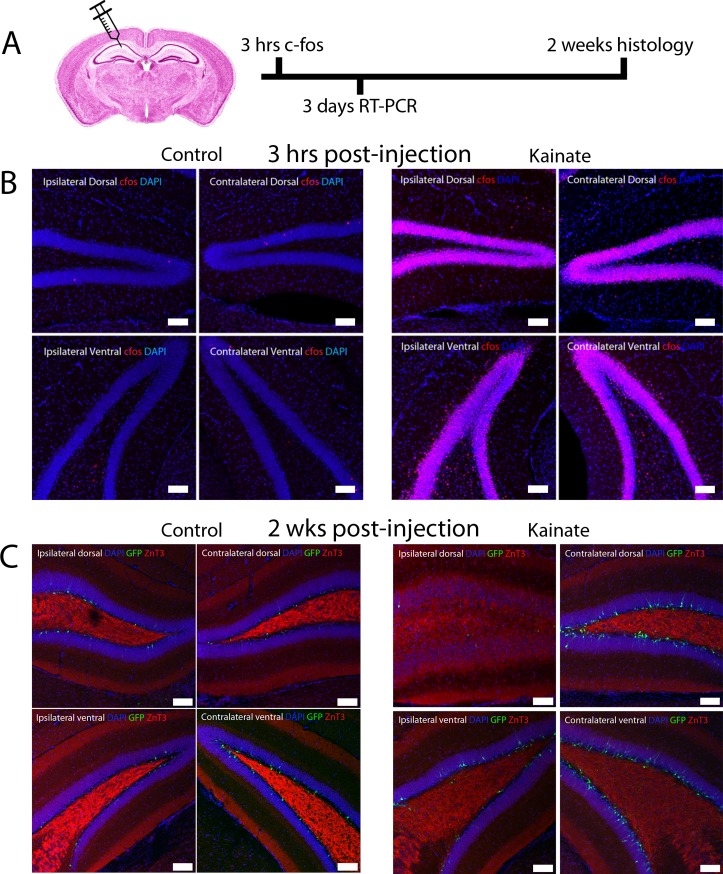
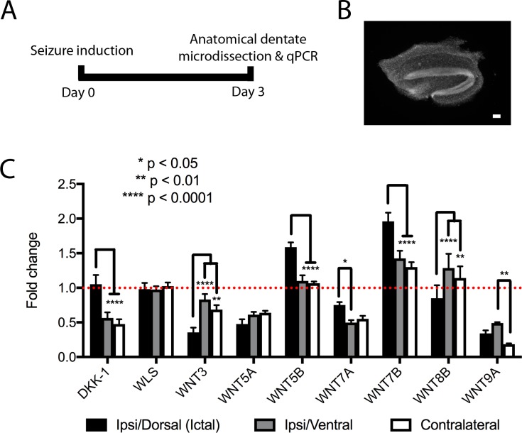
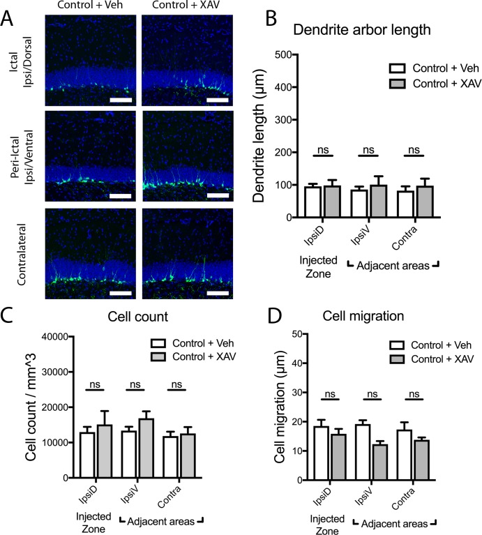
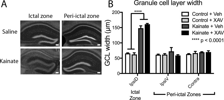
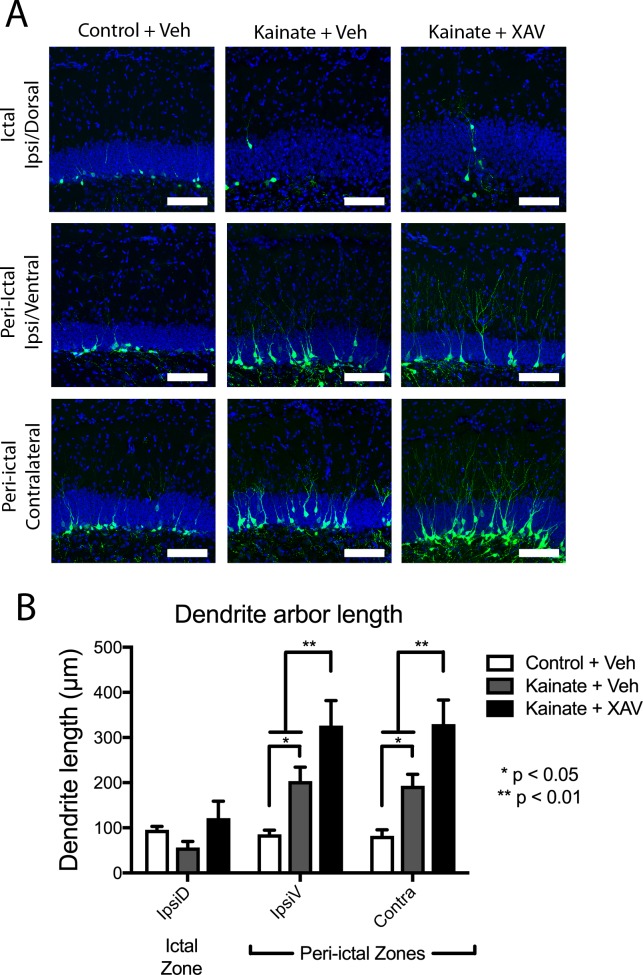
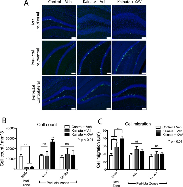
Mouse models of mesial temporal lobe epilepsy recapitulate aspects of human epilepsy, which is characterized by neuronal network remodeling in the hippocampal dentate gyrus. Observational studies suggest that this remodeling is associated with altered Wnt pathway signaling, although this has not been experimentally examined. We used the well-characterized mouse intrahippocampal kainate model of temporal lobe epilepsy to examine associations between hippocampal neurogenesis and altered Wnt signaling after seizure induction. Tissue was analyzed using immunohistochemistry and confocal microscopy, and gene expression analysis was performed by RT-qPCR on RNA extracted from anatomically micro-dissected dentate gyri. Seizures increased neurogenesis and dendritic arborization of newborn hippocampal dentate granule cells in peri-ictal regions, and decreased neurogenesis in the ictal zone, 2-weeks after kainate injection. Interestingly, administration of the novel canonical Wnt pathway inhibitor XAV939 daily for 2-weeks after kainate injection further increased dendritic arborization in peri-ictal regions after seizure, without an effect on baseline neurogenesis in control animals. Transcriptome analysis of dentate gyri demonstrated significant canonical Wnt gene dysregulation in kainate-injected mice across all regions for Wnt3, 5a and 9a. Intriguingly, certain Wnt genes demonstrated differential patterns of dysregulation between the ictal and peri-ictal zones, most notably Wnt5B, 7B and DKK-1. Together, these results demonstrate regional variation in Wnt pathway dysregulation early after seizure induction, and surprisingly, suggest that some Wnt-mediated effects might actually temper aberrant neurogenesis after seizures. The Wnt pathway may therefore provide suitable targets for novel therapies that prevent network remodeling and the development of epileptic foci in high-risk patients.
鼠内侧颞叶癫痫模型再现了人类癫痫的某些特征,其特征在于海马齿状回神经元网络重塑。观察性研究表明,这种重塑与 Wnt 途径信号改变有关,尽管尚未对此进行实验研究。我们使用了经过充分研究的海马内海人酸癫痫模型,研究了癫痫发作后海马神经发生和 Wnt 信号改变之间的关联。使用免疫组织化学和共聚焦显微镜分析组织,并对从解剖微切割的齿状回提取的 RNA 进行 RT-qPCR 基因表达分析。癫痫发作增加了peri-ictal 区域新生海马齿状颗粒细胞的神经发生和树突分支,而在海人酸注射后 2 周,ictal 区的神经发生减少。有趣的是,在海人酸注射后 2 周内每天给予新型经典 Wnt 途径抑制剂 XAV939 可进一步增加癫痫发作后 peri-ictal 区域的树突分支,而对对照动物的基线神经发生没有影响。齿状回的转录组分析表明,在所有区域中,Wnt3、5a 和 9a 的经典 Wnt 基因在海人酸注射小鼠中均存在显著失调。有趣的是,某些 Wnt 基因在 ictal 和 peri-ictal 区之间表现出不同的失调模式,最显著的是 Wnt5B、7B 和 DKK-1。总之,这些结果表明,在癫痫发作后早期,Wnt 途径失调存在区域性差异,令人惊讶的是,这表明某些 Wnt 介导的效应实际上可能抑制癫痫发作后的异常神经发生。因此,Wnt 途径可能为预防高危患者网络重塑和癫痫灶发展的新型治疗方法提供合适的靶点。