Department of Pharmacology, The School of Pharmacy, Fujian Medical University, Fuzhou, P.R. China.
Fujian Provincial Key Laboratory of Natural Medicine Pharmacology, Fuzhou, P.R. China.
Biosci Rep. 2019 Nov 29;39(11). doi: 10.1042/BSR20190817.
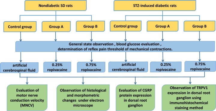
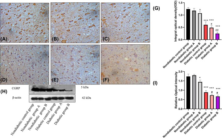
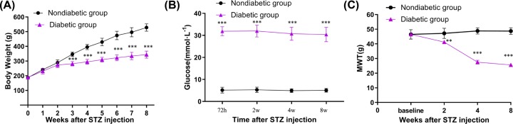
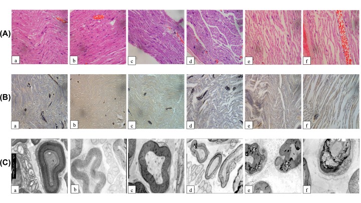
Objective To determine the effect of ropivacaine on peripheral neuropathy in diabetic rats and its possible mechanism. Methods Forty-eight Sprague-Dawley rats were randomly divided into six groups: nondiabetic control group, nondiabetic group A (0.25% ropivacaine), nondiabetic group B (0.75% ropivacaine), diabetic control group (diabetic peripheral neuropathy (DPN) +artificial cerebrospinal fluid), diabetic group A (DPN+0.25% ropivacaine), and diabetic group B (DPN + 0.75% ropivacaine), with eight rats in each group. Within an hour of the last administration, the sciatic motor nerve conduction velocity (MNCV) of each group was measured, and the morphological changes of rat sciatic nerve were observed by HE, Weil's staining and electron microscopy. The expression of transient receptor potential vanilloid (TRPV1) in the spinal cord dorsal horn of rats was analyzed by immunohistochemistry, and the expression of Calcitonin gene-related peptide (CGRP) protein in the spinal cord was analyzed by Western blot. Results Compared with the nondiabetic control group, elevated blood glucose, decreased weight and reduced average mechanical withdrawal threshold (MWT), additionally, the sciatic nerves showed significantly slowed conduction velocity (both P<0.001) and damaged pathological structure, the expression of TRPV1 and CGRP were decreased (both P<0.001) in the diabetic groups. Compared with the diabetic control group, down-regulation of TRPV1 and CGRP in spinal cord was significant for the diabetic groups A and B treated with 0.25 and 0.75% ropivacaine, the higher concentration of ropivacaine correlated with a greater change. Conclusion Ropivacaine can significantly block sciatic nerve conduction velocity in DPN rats in a concentration-dependent manner, which may be related to the expression of the TRPV1-CGRP pathway.
目的 观察罗哌卡因对糖尿病大鼠周围神经病变的影响及其可能机制。方法 将 48 只 SD 大鼠随机分为 6 组:正常对照组、非糖尿病组 A(0.25%罗哌卡因)、非糖尿病组 B(0.75%罗哌卡因)、糖尿病对照组(糖尿病周围神经病变+人工脑脊液)、糖尿病组 A(糖尿病周围神经病变+0.25%罗哌卡因)和糖尿病组 B(糖尿病周围神经病变+0.75%罗哌卡因),每组 8 只。末次给药 1 h 后,检测各组大鼠坐骨神经运动神经传导速度(MNCV),HE、Weil 染色和电镜观察大鼠坐骨神经形态学变化,免疫组化法分析大鼠脊髓背角中瞬时受体电位香草酸亚型 1(TRPV1)的表达,Western blot 法分析脊髓中降钙素基因相关肽(CGRP)蛋白的表达。结果 与正常对照组比较,糖尿病组大鼠血糖升高,体质量降低,平均机械缩足阈值降低,坐骨神经 MNCV 明显减慢(均 P<0.001),病理结构损伤明显;脊髓背角 TRPV1 和 CGRP 表达均降低(均 P<0.001)。与糖尿病对照组比较,糖尿病组 A 和 B 中 0.25%和 0.75%罗哌卡因治疗组大鼠脊髓背角中 TRPV1 和 CGRP 表达下调,高浓度罗哌卡因组下调更明显。结论 罗哌卡因可浓度依赖性显著阻滞糖尿病大鼠坐骨神经传导速度,其作用机制可能与 TRPV1-CGRP 通路的表达有关。