Xu Jian, Piao Hulin, Wang Duo, Wang Yong, Li Bo, Wang Tiance, Zhu Zhicheng, Li Dan, Xu Rihao, Liu Kexiang
Department of Cardiovascular Surgery, Second Hospital of Bethune, Jilin University, No. 218 Ziqiang Street, Changchun, 130022, China.
J Cardiothorac Surg. 2019 Nov 15;14(1):199. doi: 10.1186/s13019-019-1005-9.
Cardiac cavernous hemangiomas are extremely rare and usually difficult to be diagnosed for being asymptomatic.
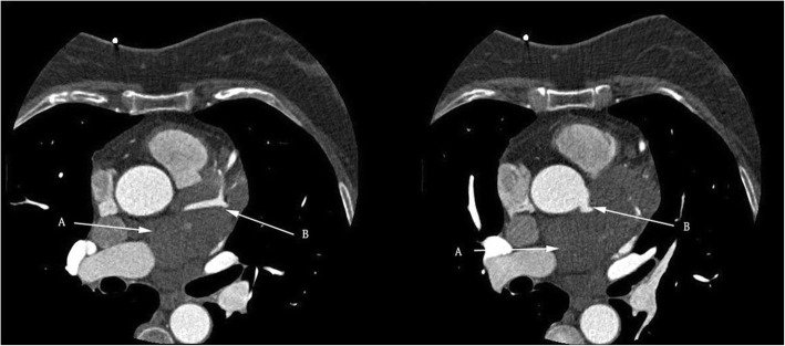
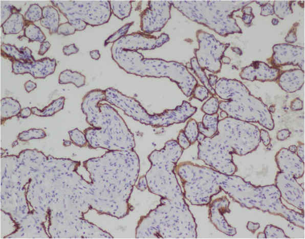
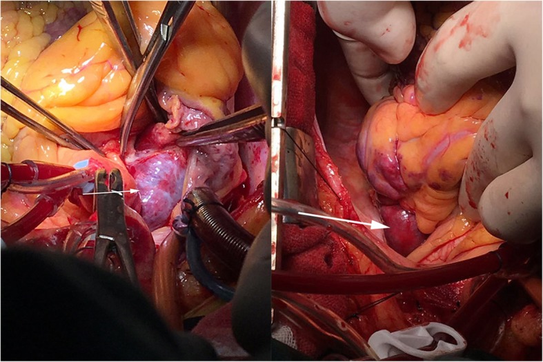
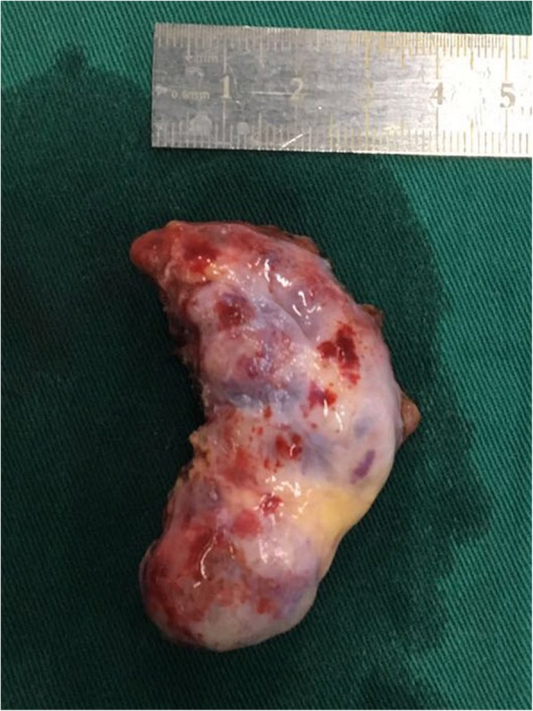
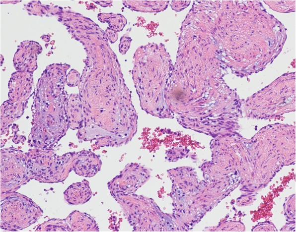
An asymptomatic 56-year-old woman was hospitalized due to a heart mass found by chest computed tomography (CT) during her annual physical examination. Coronary computed tomography angiography (CTA) disclosed a tumorous lesion, located in the left atrial roof and extended to the posterior wall of the aortic root and surrounding the left main coronary artery. However, there was no communicating branches between the hemangioma and coronary artery and no coronary artery stenosis. The tumor was excised with low-frequency electrocautery under cardiopulmonary bypass. The histopathological examination indicated the mass a cavernous hemangioma. The patient was discharged with an uneventful recovery.
Here we presented a rare case of successfully excision of a cavernous hemangioma involving the left atrial roof and left coronary artery. We advocate adequate exposure and complete surgical excision with low-frequency electrocautery to avoid remnants and excessive resection.
心脏海绵状血管瘤极为罕见,通常因无症状而难以诊断。
一名56岁无症状女性因年度体检时胸部计算机断层扫描(CT)发现心脏肿物而住院。冠状动脉计算机断层扫描血管造影(CTA)显示一个肿瘤性病变,位于左心房顶部,延伸至主动脉根部后壁并包绕左冠状动脉主干。然而,血管瘤与冠状动脉之间无交通支,也无冠状动脉狭窄。在体外循环下用低频电灼切除肿瘤。组织病理学检查显示肿物为海绵状血管瘤。患者康复顺利出院。
我们在此报告了一例成功切除累及左心房顶部和左冠状动脉的海绵状血管瘤的罕见病例。我们主张充分暴露并采用低频电灼进行完整手术切除,以避免残留和过度切除。