• 文献检索
  • 文档翻译
  • 深度研究
  • 学术资讯
  • Suppr Zotero 插件Zotero 插件
  • 邀请有礼
  • 套餐&价格
  • 历史记录
应用&插件
Suppr Zotero 插件Zotero 插件浏览器插件Mac 客户端Windows 客户端微信小程序
定价
高级版会员购买积分包购买API积分包
服务
文献检索文档翻译深度研究API 文档MCP 服务
关于我们
关于 Suppr公司介绍联系我们用户协议隐私条款
关注我们

Suppr 超能文献

核心技术专利:CN118964589B侵权必究
粤ICP备2023148730 号-1Suppr @ 2026

文献检索

告别复杂PubMed语法,用中文像聊天一样搜索,搜遍4000万医学文献。AI智能推荐,让科研检索更轻松。

立即免费搜索

文件翻译

保留排版,准确专业,支持PDF/Word/PPT等文件格式,支持 12+语言互译。

免费翻译文档

深度研究

AI帮你快速写综述,25分钟生成高质量综述,智能提取关键信息,辅助科研写作。

立即免费体验

溶酶体的组装和拆解通过利什曼原虫的细胞周期改变内吞作用的速率。

Lysosome assembly and disassembly changes endocytosis rate through the Leishmania cell cycle.

机构信息

Sir William Dunn School of Pathology, University of Oxford, Oxford, UK.

The Peter Medawar Building for Pathogen Research, University of Oxford, Oxford, UK.

出版信息

Microbiologyopen. 2020 Feb;9(2):e969. doi: 10.1002/mbo3.969. Epub 2019 Nov 19.

DOI:10.1002/mbo3.969
PMID:31743959
原文链接:https://pmc.ncbi.nlm.nih.gov/articles/PMC7002101/
Abstract

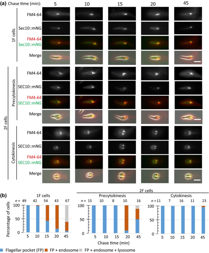
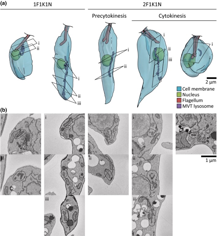
The Leishmania lysosome has an atypical structure, consisting of an elongated vesicle-filled tubule running along the anterior-posterior axis of the cell, which is termed the multivesicular tubule (MVT) lysosome. Alongside, the MVT lysosome is one or more microtubules, the lysosomal microtubule(s). Previous work indicated there were cell cycle-related changes in MVT lysosome organization; however, these only provided snapshots and did not connect the changes in the lysosomal microtubule(s) or lysosomal function. Using mNeonGreen tagged cysteine peptidase A and SPEF1 as markers of the MVT lysosome and lysosomal microtubule(s), we examined the dynamics of these structures through the cell cycle. Both the MVT lysosome and lysosomal microtubule(s) elongated at the beginning of the cell cycle before plateauing and then disassembling in late G before cytokinesis. Moreover, the endocytic rate in cells where the MVT lysosome and lysosomal microtubule(s) had disassembled was extremely low. The dynamic nature of the MVT lysosome and lysosomal microtubule(s) parallels that of the Trypanosoma cruzi cytostome/cytopharynx, which also has a similar membrane tubule structure with associated microtubules. As the cytostome/cytopharynx is an ancestral feature of the kinetoplastids, this suggests that the Leishmania MVT lysosome and lysosomal microtubule(s) are a reduced cytostome/cytopharynx-like feature.

摘要

利什曼虫溶酶体具有非典型的结构,由一个沿细胞前后轴延伸的充满囊泡的管状结构组成,称为多泡管状溶酶体(MVT)溶酶体。此外,MVT 溶酶体还有一个或多个微管,即溶酶体微管。先前的工作表明,MVT 溶酶体的结构在细胞周期中发生相关变化;然而,这些变化仅提供了静态快照,并未将溶酶体微管或溶酶体功能的变化联系起来。我们使用标记有 mNeonGreen 的半胱氨酸肽酶 A 和 SPEF1 作为 MVT 溶酶体和溶酶体微管的标志物,通过细胞周期来研究这些结构的动态变化。在细胞周期开始时,MVT 溶酶体和溶酶体微管均伸长,然后在后期 G 时达到平台期并解体,最后在细胞分裂时解体。此外,在 MVT 溶酶体和溶酶体微管已经解体的细胞中,内吞作用的速率极低。MVT 溶酶体和溶酶体微管的动态性质与克氏锥虫胞口/胞咽的动态性质相似,后者也具有类似的膜管状结构和相关的微管。由于胞口/胞咽是动基体门的一个古老特征,这表明利什曼虫的 MVT 溶酶体和溶酶体微管是一种简化的胞口/胞咽样特征。

https://cdn.ncbi.nlm.nih.gov/pmc/blobs/eb83/7002101/9cbeff80a050/MBO3-9-e969-g009.jpg
https://cdn.ncbi.nlm.nih.gov/pmc/blobs/eb83/7002101/8fc38cae49b7/MBO3-9-e969-g001.jpg
https://cdn.ncbi.nlm.nih.gov/pmc/blobs/eb83/7002101/7f49b0b8969e/MBO3-9-e969-g002.jpg
https://cdn.ncbi.nlm.nih.gov/pmc/blobs/eb83/7002101/79255f3c5477/MBO3-9-e969-g003.jpg
https://cdn.ncbi.nlm.nih.gov/pmc/blobs/eb83/7002101/e506e50c676d/MBO3-9-e969-g004.jpg
https://cdn.ncbi.nlm.nih.gov/pmc/blobs/eb83/7002101/07fc168ad2ca/MBO3-9-e969-g005.jpg
https://cdn.ncbi.nlm.nih.gov/pmc/blobs/eb83/7002101/32e7d5ce98a4/MBO3-9-e969-g006.jpg
https://cdn.ncbi.nlm.nih.gov/pmc/blobs/eb83/7002101/0f3f402fad8d/MBO3-9-e969-g007.jpg
https://cdn.ncbi.nlm.nih.gov/pmc/blobs/eb83/7002101/40b7099958b0/MBO3-9-e969-g008.jpg
https://cdn.ncbi.nlm.nih.gov/pmc/blobs/eb83/7002101/9cbeff80a050/MBO3-9-e969-g009.jpg

相似文献

1
Lysosome assembly and disassembly changes endocytosis rate through the Leishmania cell cycle.溶酶体的组装和拆解通过利什曼原虫的细胞周期改变内吞作用的速率。
Microbiologyopen. 2020 Feb;9(2):e969. doi: 10.1002/mbo3.969. Epub 2019 Nov 19.
2
The cytostome-cytopharynx complex of Trypanosoma cruzi epimastigotes disassembles during cell division.克氏锥虫上鞭毛体的胞口-胞咽复合体在细胞分裂过程中会解体。
J Cell Sci. 2017 Jan 1;130(1):164-176. doi: 10.1242/jcs.187419. Epub 2016 Jun 30.
3
The three-dimensional structure of the cytostome-cytopharynx complex of Trypanosoma cruzi epimastigotes.克氏锥虫上鞭毛体胞口-胞咽复合体的三维结构。
J Cell Sci. 2014 May 15;127(Pt 10):2227-37. doi: 10.1242/jcs.135491. Epub 2014 Mar 7.
4
Loss of the cytostome-cytopharynx and endocytic ability are late events in Trypanosoma cruzi metacyclogenesis.胞口-胞咽的丧失和内吞能力是克氏锥虫循环后期的事件。
J Struct Biol. 2016 Dec;196(3):319-328. doi: 10.1016/j.jsb.2016.07.018. Epub 2016 Jul 30.
5
Ultrastructural and cytochemical identification of megasome in Leishmania (Leishmania) chagasi.杜氏利什曼原虫(利什曼原虫属)中巨大颗粒的超微结构和细胞化学鉴定
Parasitol Res. 2004 Feb;92(3):246-54. doi: 10.1007/s00436-003-1027-y. Epub 2004 Jan 9.
6
Regulated degradation of an endoplasmic reticulum membrane protein in a tubular lysosome in Leishmania mexicana.墨西哥利什曼原虫内质网膜蛋白在管状溶酶体中的调控降解
Mol Biol Cell. 2001 Aug;12(8):2364-77. doi: 10.1091/mbc.12.8.2364.
7
Dynamics of the orphan myosin MyoF over Trypanosoma cruzi life cycle and along the endocytic pathway.孤儿肌球蛋白 MyoF 在克氏锥虫生活史和胞吞途径中的动态变化。
Parasitol Int. 2022 Feb;86:102444. doi: 10.1016/j.parint.2021.102444. Epub 2021 Aug 28.
8
Developmental changes in lysosome morphology and function Leishmania parasites.利什曼原虫溶酶体形态和功能的发育变化
Int J Parasitol. 2002 Nov;32(12):1435-45. doi: 10.1016/s0020-7519(02)00140-6.
9
The cytostome-cytopharynx complex of intracellular and extracellular amastigotes of Trypanosoma cruzi exhibit structural and functional differences.克氏锥虫细胞内和细胞外无鞭毛体的胞口-胞咽复合体表现出结构和功能上的差异。
Cell Microbiol. 2021 Sep;23(9):e13346. doi: 10.1111/cmi.13346. Epub 2021 May 18.
10
Role for the flagellum attachment zone in Leishmania anterior cell tip morphogenesis.鞭毛附着区在利什曼原虫前体细胞顶端形态发生中的作用。
PLoS Pathog. 2020 Oct 22;16(10):e1008494. doi: 10.1371/journal.ppat.1008494. eCollection 2020 Oct.

引用本文的文献

1
Tubulin detyrosination shapes cytoskeletal architecture and virulence.微管蛋白去酪氨酸化塑造细胞骨架结构和毒力。
Proc Natl Acad Sci U S A. 2025 Jan 21;122(3):e2415296122. doi: 10.1073/pnas.2415296122. Epub 2025 Jan 14.
2
Whole cell reconstructions of Leishmania mexicana through the cell cycle.墨西哥利什曼原虫整个细胞周期的重建
PLoS Pathog. 2024 Feb 28;20(2):e1012054. doi: 10.1371/journal.ppat.1012054. eCollection 2024 Feb.
3
Spef1/CLAMP binds microtubules and actin-based structures and regulates cell migration and epithelia cell polarity.

本文引用的文献

1
flagellum attachment zone is critical for flagellar pocket shape, development in the sand fly, and pathogenicity in the host.鞭毛附着区对于鞭毛囊的形状、在沙蝇中的发育以及在宿主中的致病性至关重要。
Proc Natl Acad Sci U S A. 2019 Mar 26;116(13):6351-6360. doi: 10.1073/pnas.1812462116. Epub 2019 Mar 8.
2
Cellular landmarks of Trypanosoma brucei and Leishmania mexicana.布氏锥虫和墨西哥利什曼原虫的细胞标志物
Mol Biochem Parasitol. 2019 Jun;230:24-36. doi: 10.1016/j.molbiopara.2018.12.003. Epub 2018 Dec 11.
3
ImageJ2: ImageJ for the next generation of scientific image data.
Spef1/CLAMP 结合微管和肌动蛋白为基础的结构,调节细胞迁移和上皮细胞极性。
Ann N Y Acad Sci. 2022 Sep;1515(1):97-104. doi: 10.1111/nyas.14845. Epub 2022 Jun 16.
4
The kinesin of the flagellum attachment zone in Leishmania is required for cell morphogenesis, cell division and virulence in the mammalian host.鞭毛连接区域的驱动蛋白在利什曼原虫的细胞形态发生、细胞分裂和在哺乳动物宿主体内的毒力中是必需的。
PLoS Pathog. 2021 Jun 18;17(6):e1009666. doi: 10.1371/journal.ppat.1009666. eCollection 2021 Jun.
5
The Gene Encodes for an Atypical Dual Specificity Lipid-Like Phosphatase Expressed in Promastigotes and Amastigotes; Substrate Specificity, Intracellular Localizations, and Putative Role(s).该基因编码一种在前鞭毛体和无鞭毛体中表达的非典型双特异性脂质样磷酸酶;底物特异性、细胞内定位和潜在作用。
Front Cell Infect Microbiol. 2021 Mar 25;11:591868. doi: 10.3389/fcimb.2021.591868. eCollection 2021.
6
The single flagellum of has a fixed polarisation of its asymmetric beat.具有固定极性的单一鞭毛呈不对称拍打。
J Cell Sci. 2020 Oct 22;133(20):jcs246637. doi: 10.1242/jcs.246637.
7
Basal Body Protein TbSAF1 Is Required for Microtubule Quartet Anchorage to the Basal Bodies in .TbSAF1 基体质蛋白对于微管四联体锚定到基体是必需的。
mBio. 2020 Jun 9;11(3):e00668-20. doi: 10.1128/mBio.00668-20.
ImageJ2:面向下一代科学图像数据的ImageJ。
BMC Bioinformatics. 2017 Nov 29;18(1):529. doi: 10.1186/s12859-017-1934-z.
4
Extensive flagellar remodeling during the complex life cycle of , an early-branching trypanosomatid.在早期分支的动基体目生物 的复杂生命周期中,广泛的鞭毛重塑。
Proc Natl Acad Sci U S A. 2017 Oct 31;114(44):11757-11762. doi: 10.1073/pnas.1712311114. Epub 2017 Oct 16.
5
Mechanisms and functions of lysosome positioning.溶酶体定位的机制与功能。
J Cell Sci. 2016 Dec 1;129(23):4329-4339. doi: 10.1242/jcs.196287. Epub 2016 Oct 31.
6
The cytostome-cytopharynx complex of Trypanosoma cruzi epimastigotes disassembles during cell division.克氏锥虫上鞭毛体的胞口-胞咽复合体在细胞分裂过程中会解体。
J Cell Sci. 2017 Jan 1;130(1):164-176. doi: 10.1242/jcs.187419. Epub 2016 Jun 30.
7
The Flagellum Attachment Zone: 'The Cellular Ruler' of Trypanosome Morphology.鞭毛附着区:锥虫形态学的“细胞标尺”
Trends Parasitol. 2016 Apr;32(4):309-324. doi: 10.1016/j.pt.2015.12.010. Epub 2016 Jan 8.
8
Flagellar pocket restructuring through the Leishmania life cycle involves a discrete flagellum attachment zone.利什曼原虫生活周期中鞭毛袋的重组涉及一个离散的鞭毛附着区。
J Cell Sci. 2016 Feb 15;129(4):854-67. doi: 10.1242/jcs.183152. Epub 2016 Jan 8.
9
A toolkit enabling efficient, scalable and reproducible gene tagging in trypanosomatids.一种能够在锥虫中实现高效、可扩展且可重复的基因标记的工具包。
Open Biol. 2015 Jan;5(1):140197. doi: 10.1098/rsob.140197.
10
The three-dimensional structure of the cytostome-cytopharynx complex of Trypanosoma cruzi epimastigotes.克氏锥虫上鞭毛体胞口-胞咽复合体的三维结构。
J Cell Sci. 2014 May 15;127(Pt 10):2227-37. doi: 10.1242/jcs.135491. Epub 2014 Mar 7.