• 文献检索
  • 文档翻译
  • 深度研究
  • 学术资讯
  • Suppr Zotero 插件Zotero 插件
  • 邀请有礼
  • 套餐&价格
  • 历史记录
应用&插件
Suppr Zotero 插件Zotero 插件浏览器插件Mac 客户端Windows 客户端微信小程序
定价
高级版会员购买积分包购买API积分包
服务
文献检索文档翻译深度研究API 文档MCP 服务
关于我们
关于 Suppr公司介绍联系我们用户协议隐私条款
关注我们

Suppr 超能文献

核心技术专利:CN118964589B侵权必究
粤ICP备2023148730 号-1Suppr @ 2026

文献检索

告别复杂PubMed语法,用中文像聊天一样搜索,搜遍4000万医学文献。AI智能推荐,让科研检索更轻松。

立即免费搜索

文件翻译

保留排版,准确专业,支持PDF/Word/PPT等文件格式,支持 12+语言互译。

免费翻译文档

深度研究

AI帮你快速写综述,25分钟生成高质量综述,智能提取关键信息,辅助科研写作。

立即免费体验

钾离子通道 Kcne3 是一种血管内皮细胞尖端细胞选择性表达的 VEGFA 诱导基因。

The potassium channel Kcne3 is a VEGFA-inducible gene selectively expressed by vascular endothelial tip cells.

机构信息

Department of Pre-Therapeutic Target Discovery, Regeneron Pharmaceuticals Inc, Tarrytown, NY, 10591, USA.

Palmira Biopharma, Moscow, 143026, RF, Russia.

出版信息

Angiogenesis. 2020 May;23(2):179-192. doi: 10.1007/s10456-019-09696-8. Epub 2019 Nov 21.

DOI:10.1007/s10456-019-09696-8
PMID:31754927
原文链接:https://pmc.ncbi.nlm.nih.gov/articles/PMC7160073/
Abstract

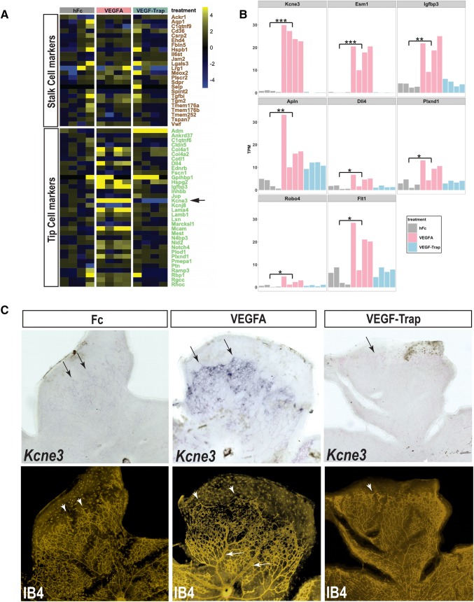
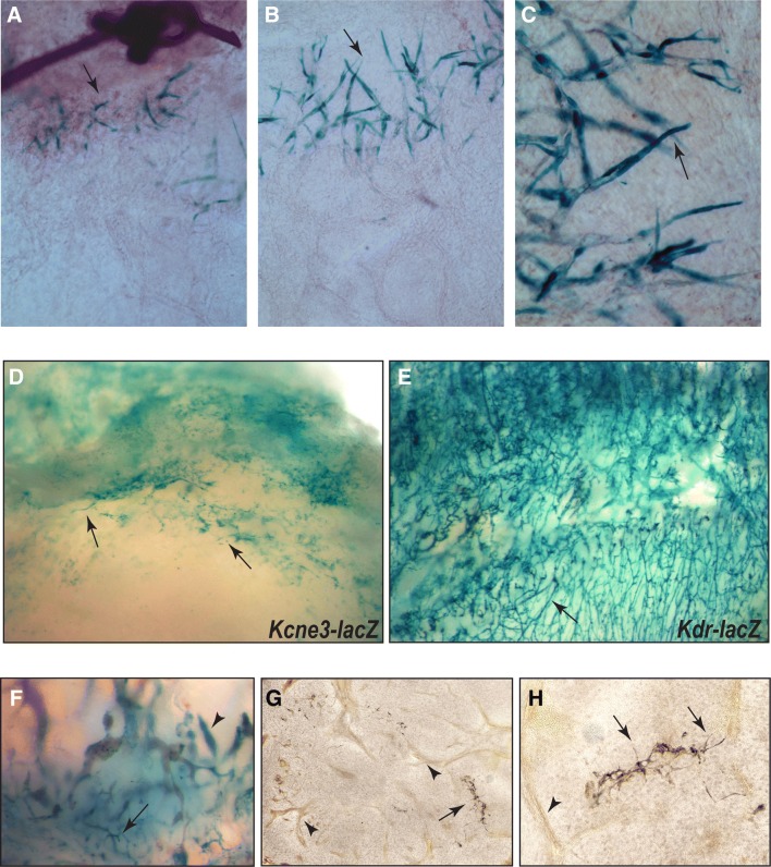
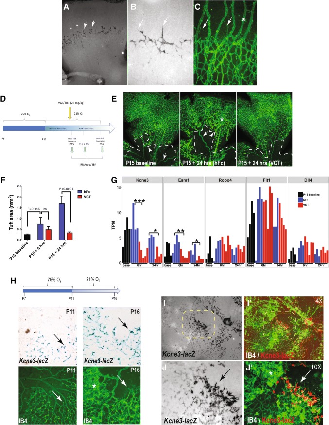
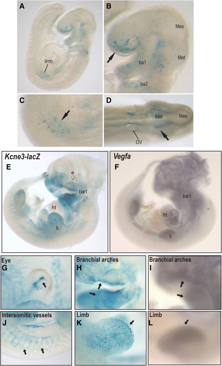
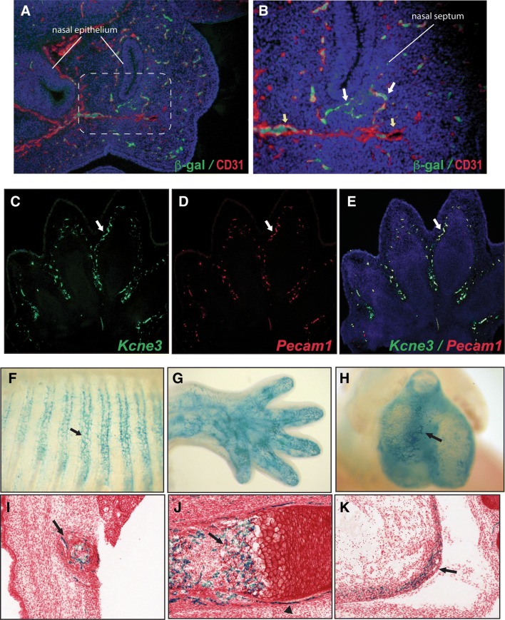
Angiogenesis is largely driven by motile endothelial tip-cells capable of invading avascular tissue domains and enabling new vessel formation. Highly responsive to Vascular Endothelial Growth-Factor-A (VEGFA), endothelial tip-cells also suppress angiogenic sprouting in adjacent stalk cells, and thus have been a primary therapeutic focus in addressing neovascular pathologies. Surprisingly, however, there remains a paucity of specific endothelial tip-cell markers. Here, we employ transcriptional profiling and a lacZ reporter allele to identify Kcne3 as an early and selective endothelial tip-cell marker in multiple angiogenic contexts. In development, Kcne3 expression initiates during early phases of angiogenesis (E9) and remains specific to endothelial tip-cells, often adjacent to regions expressing VEGFA. Consistently, Kcne3 activation is highly responsive to exogenous VEGFA but maintains tip-cell specificity throughout normal retinal angiogenesis. We also demonstrate endothelial tip-cell selectivity of Kcne3 in several injury and tumor models. Together, our data show that Kcne3 is a unique marker of sprouting angiogenic tip-cells and offers new opportunities for investigating and targeting this cell type.

摘要

血管生成在很大程度上是由能够侵袭无血管组织区域并促进新血管形成的运动性内皮尖端细胞驱动的。内皮尖端细胞对血管内皮生长因子-A(VEGFA)高度敏感,还能抑制相邻的茎细胞中的血管生成芽生,因此一直是解决新血管病理的主要治疗重点。然而,令人惊讶的是,特异性内皮尖端细胞标志物仍然很少。在这里,我们利用转录谱分析和一个 lacZ 报告基因等位基因,鉴定出 Kcne3 是多种血管生成情况下早期和选择性的内皮尖端细胞标志物。在发育过程中,Kcne3 的表达在血管生成的早期阶段(E9)开始,并始终特异性地表达于内皮尖端细胞,通常与表达 VEGFA 的区域相邻。一致地,Kcne3 的激活对外源性 VEGFA 高度敏感,但在正常视网膜血管生成过程中始终保持尖端细胞的特异性。我们还在几种损伤和肿瘤模型中证明了 Kcne3 的内皮尖端细胞选择性。总之,我们的数据表明,Kcne3 是发芽血管生成尖端细胞的独特标志物,为研究和靶向这种细胞类型提供了新的机会。

https://cdn.ncbi.nlm.nih.gov/pmc/blobs/2176/7160073/f6f0146e537d/10456_2019_9696_Fig5_HTML.jpg
https://cdn.ncbi.nlm.nih.gov/pmc/blobs/2176/7160073/68c984b24714/10456_2019_9696_Fig1_HTML.jpg
https://cdn.ncbi.nlm.nih.gov/pmc/blobs/2176/7160073/3a0b8b788922/10456_2019_9696_Fig2_HTML.jpg
https://cdn.ncbi.nlm.nih.gov/pmc/blobs/2176/7160073/e1a3db4c534f/10456_2019_9696_Fig3_HTML.jpg
https://cdn.ncbi.nlm.nih.gov/pmc/blobs/2176/7160073/194961e1a013/10456_2019_9696_Fig4_HTML.jpg
https://cdn.ncbi.nlm.nih.gov/pmc/blobs/2176/7160073/f6f0146e537d/10456_2019_9696_Fig5_HTML.jpg

相似文献

1
The potassium channel Kcne3 is a VEGFA-inducible gene selectively expressed by vascular endothelial tip cells.钾离子通道 Kcne3 是一种血管内皮细胞尖端细胞选择性表达的 VEGFA 诱导基因。
Angiogenesis. 2020 May;23(2):179-192. doi: 10.1007/s10456-019-09696-8. Epub 2019 Nov 21.
2
Notch pathway targets proangiogenic regulator Sox17 to restrict angiogenesis.Notch 通路靶向促血管生成调节因子 Sox17 以限制血管生成。
Circ Res. 2014 Jul 7;115(2):215-26. doi: 10.1161/CIRCRESAHA.115.303142. Epub 2014 Apr 22.
3
MEF2 transcription factors are key regulators of sprouting angiogenesis.MEF2转录因子是出芽血管生成的关键调节因子。
Genes Dev. 2016 Oct 15;30(20):2297-2309. doi: 10.1101/gad.290619.116.
4
Endothelial-specific YY1 governs sprouting angiogenesis through directly interacting with RBPJ.内皮细胞特异性 YY1 通过直接与 RBPJ 相互作用来调控血管生成。
Proc Natl Acad Sci U S A. 2020 Mar 3;117(9):4792-4801. doi: 10.1073/pnas.1916198117. Epub 2020 Feb 19.
5
Retinal myeloid cells regulate tip cell selection and vascular branching morphogenesis via Notch ligand Delta-like 1.视网膜髓样细胞通过 Notch 配体 Delta-like 1 调节尖端细胞选择和血管分支形态发生。
Sci Rep. 2019 Jul 5;9(1):9798. doi: 10.1038/s41598-019-46308-3.
6
NRP1 acts cell autonomously in endothelium to promote tip cell function during sprouting angiogenesis.NRP1 在血管生成过程中自主作用于内皮细胞,促进尖端细胞的功能。
Blood. 2013 Mar 21;121(12):2352-62. doi: 10.1182/blood-2012-05-424713. Epub 2013 Jan 11.
7
eNOS controls angiogenic sprouting and retinal neovascularization through the regulation of endothelial cell polarity.内皮型一氧化氮合酶通过调节内皮细胞极性控制血管生成芽生和视网膜新生血管形成。
Cell Mol Life Sci. 2021 Dec 31;79(1):37. doi: 10.1007/s00018-021-04042-y.
8
Extrinsic Notch ligand Delta-like 1 regulates tip cell selection and vascular branching morphogenesis.外在 Notch 配体 Delta-like 1 调节尖端细胞选择和血管分支形态发生。
Circ Res. 2012 Feb 17;110(4):530-5. doi: 10.1161/CIRCRESAHA.111.263319. Epub 2012 Jan 26.
9
CD34 Promotes Pathological Epi-Retinal Neovascularization in a Mouse Model of Oxygen-Induced Retinopathy.CD34在氧诱导性视网膜病变小鼠模型中促进病理性视网膜前新生血管形成。
PLoS One. 2016 Jun 28;11(6):e0157902. doi: 10.1371/journal.pone.0157902. eCollection 2016.
10
VEGF-A/Notch-Induced Podosomes Proteolyse Basement Membrane Collagen-IV during Retinal Sprouting Angiogenesis.VEGF-A/Notch诱导的足体在视网膜新生血管形成过程中降解基底膜胶原蛋白IV
Cell Rep. 2016 Oct 4;17(2):484-500. doi: 10.1016/j.celrep.2016.09.016.

引用本文的文献

1
Identification of New Markers of Angiogenic Sprouting Using Transcriptomics: New Role for RND3.利用转录组学鉴定血管生成芽生的新标志物:RND3 的新作用。
Arterioscler Thromb Vasc Biol. 2024 May;44(5):e145-e167. doi: 10.1161/ATVBAHA.123.320599. Epub 2024 Mar 14.
2
Succinate-induced macrophage polarization and RBP4 secretion promote vascular sprouting in ocular neovascularization.琥珀酸诱导的巨噬细胞极化和 RBP4 分泌促进眼部新生血管中的血管生成。
J Neuroinflammation. 2023 Dec 21;20(1):308. doi: 10.1186/s12974-023-02998-1.
3
The role and mechanisms of microvascular damage in the ischemic myocardium.

本文引用的文献

1
Shear-Stress Sensitive Inwardly-Rectifying K Channels Regulate Developmental Retinal Angiogenesis by Vessel Regression.剪切应力敏感内向整流钾通道通过血管消退调节视网膜发育性血管生成。
Cell Physiol Biochem. 2019;52(6):1569-1583. doi: 10.33594/000000109.
2
Consensus guidelines for the use and interpretation of angiogenesis assays.血管生成分析检测应用和解释的共识指南。
Angiogenesis. 2018 Aug;21(3):425-532. doi: 10.1007/s10456-018-9613-x.
3
Single-Cell Transcriptome Analyses Reveal Endothelial Cell Heterogeneity in Tumors and Changes following Antiangiogenic Treatment.
微血管损伤在缺血性心肌中的作用和机制。
Cell Mol Life Sci. 2023 Oct 29;80(11):341. doi: 10.1007/s00018-023-04998-z.
4
Targeting FSCN1 with an oral small-molecule inhibitor for treating ocular neovascularization.靶向 FSCN1 的口服小分子抑制剂治疗眼部血管新生。
J Transl Med. 2023 Aug 18;21(1):555. doi: 10.1186/s12967-023-04225-0.
5
In Vitro Mineralisation of Tissue-Engineered Cartilage Reduces Endothelial Cell Migration, Proliferation and Tube Formation.组织工程软骨的体外矿化降低了内皮细胞的迁移、增殖和管状形成。
Cells. 2023 Apr 20;12(8):1202. doi: 10.3390/cells12081202.
6
New Insight on 2D In Vitro Angiogenesis Models: All That Stretches Is Not a Tube.二维体外血管生成模型的新视角:并非所有拉伸的都是管状结构。
Cells. 2022 Oct 18;11(20):3278. doi: 10.3390/cells11203278.
7
Expanding tubular microvessels on stiff substrates with endothelial cells and pericytes from the same adult tissue.利用来自同一成年组织的内皮细胞和周细胞在刚性基质上扩展管状微血管。
J Tissue Eng. 2022 Sep 22;13:20417314221125310. doi: 10.1177/20417314221125310. eCollection 2022 Jan-Dec.
8
Mitophagy alleviates ischemia/reperfusion-induced microvascular damage through improving mitochondrial quality control.自噬减轻缺血/再灌注诱导的微血管损伤,通过改善线粒体质量控制。
Bioengineered. 2022 Feb;13(2):3596-3607. doi: 10.1080/21655979.2022.2027065.
9
Melatonin Attenuates Ischemia/Reperfusion-Induced Oxidative Stress by Activating Mitochondrial Fusion in Cardiomyocytes.褪黑素通过激活心肌细胞中线粒体融合来减轻缺血/再灌注诱导的氧化应激。
Oxid Med Cell Longev. 2022 Jan 10;2022:7105181. doi: 10.1155/2022/7105181. eCollection 2022.
10
TMEM60 Promotes the Proliferation and Migration and Inhibits the Apoptosis of Glioma through Modulating AKT Signaling.跨膜蛋白60通过调节AKT信号通路促进胶质瘤的增殖和迁移并抑制其凋亡。
J Oncol. 2022 Jan 3;2022:9913700. doi: 10.1155/2022/9913700. eCollection 2022.
单细胞转录组分析揭示了肿瘤中的内皮细胞异质性以及抗血管生成治疗后的变化。
Cancer Res. 2018 May 1;78(9):2370-2382. doi: 10.1158/0008-5472.CAN-17-2728. Epub 2018 Feb 15.
4
VEGF amplifies transcription through ETS1 acetylation to enable angiogenesis.血管内皮生长因子通过ETS1乙酰化增强转录以促进血管生成。
Nat Commun. 2017 Aug 29;8(1):383. doi: 10.1038/s41467-017-00405-x.
5
Diabetic retinopathy: current understanding, mechanisms, and treatment strategies.糖尿病视网膜病变:当前的认识、机制及治疗策略
JCI Insight. 2017 Jul 20;2(14). doi: 10.1172/jci.insight.93751.
6
Dll4 and Notch signalling couples sprouting angiogenesis and artery formation.Dll4 和 Notch 信号通路耦连出芽型血管生成和动脉形成。
Nat Cell Biol. 2017 Aug;19(8):915-927. doi: 10.1038/ncb3555. Epub 2017 Jul 17.
7
SoxF Transcription Factors Are Positive Feedback Regulators of VEGF Signaling.SoxF 转录因子是 VEGF 信号的正反馈调节因子。
Circ Res. 2016 Sep 16;119(7):839-52. doi: 10.1161/CIRCRESAHA.116.308483. Epub 2016 Aug 15.
8
Revisiting the mouse model of oxygen-induced retinopathy.重新审视氧诱导视网膜病变的小鼠模型。
Eye Brain. 2016;8:67-79. doi: 10.2147/EB.S94447. Epub 2016 May 20.
9
KCNE1 and KCNE3: The yin and yang of voltage-gated K(+) channel regulation.KCNE1与KCNE3:电压门控钾离子通道调节中的阴阳平衡
Gene. 2016 Jan 15;576(1 Pt 1):1-13. doi: 10.1016/j.gene.2015.09.059. Epub 2015 Sep 26.
10
Arteries are formed by vein-derived endothelial tip cells.动脉由源自静脉的内皮尖端细胞形成。
Nat Commun. 2014 Dec 15;5:5758. doi: 10.1038/ncomms6758.