• 文献检索
  • 文档翻译
  • 深度研究
  • 学术资讯
  • Suppr Zotero 插件Zotero 插件
  • 邀请有礼
  • 套餐&价格
  • 历史记录
应用&插件
Suppr Zotero 插件Zotero 插件浏览器插件Mac 客户端Windows 客户端微信小程序
定价
高级版会员购买积分包购买API积分包
服务
文献检索文档翻译深度研究API 文档MCP 服务
关于我们
关于 Suppr公司介绍联系我们用户协议隐私条款
关注我们

Suppr 超能文献

核心技术专利:CN118964589B侵权必究
粤ICP备2023148730 号-1Suppr @ 2026

文献检索

告别复杂PubMed语法,用中文像聊天一样搜索,搜遍4000万医学文献。AI智能推荐,让科研检索更轻松。

立即免费搜索

文件翻译

保留排版,准确专业,支持PDF/Word/PPT等文件格式,支持 12+语言互译。

免费翻译文档

深度研究

AI帮你快速写综述,25分钟生成高质量综述,智能提取关键信息,辅助科研写作。

立即免费体验

滤泡性胰腺炎:一种罕见的胰腺炎性假瘤。

Follicular pancreatitis: A rare pancreatic inflammatory pseudotumor.

机构信息

Department of Radiology, University of California San Diego, San Diego, CA, USA.

Department of Pathology, University of California San Diego, San Diego, CA, USA; Department of Pathology, VA San Diego Healthcare System, San Diego, CA, USA.

出版信息

Clin Imaging. 2020 Jan;59(1):39-44. doi: 10.1016/j.clinimag.2019.08.006. Epub 2019 Oct 25.

DOI:10.1016/j.clinimag.2019.08.006
PMID:31756593
原文链接:https://pmc.ncbi.nlm.nih.gov/articles/PMC6938461/
Abstract

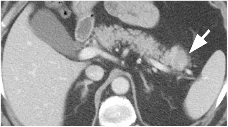
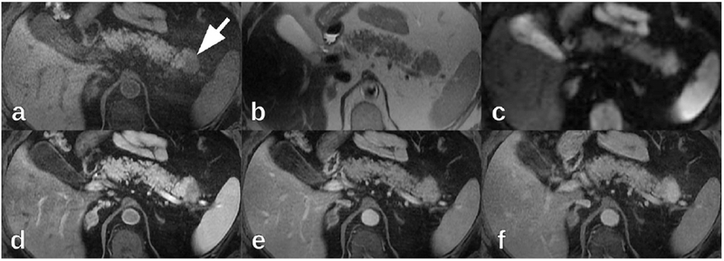
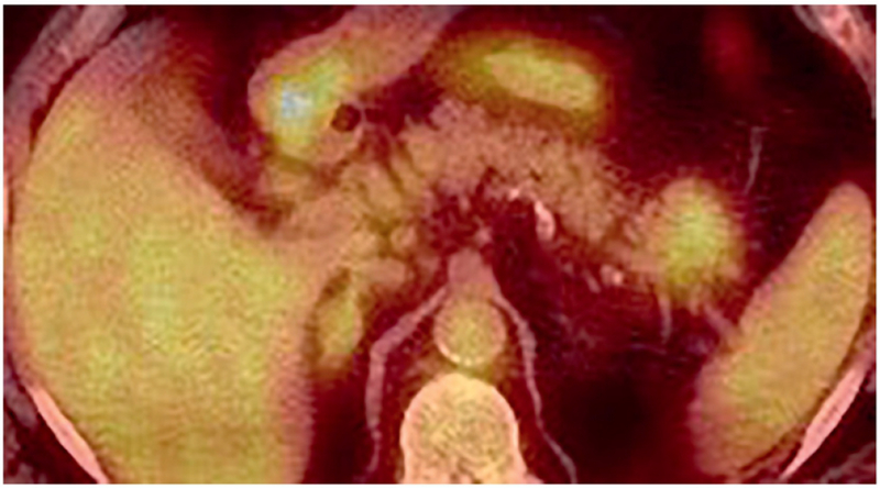
Inflammatory pseudotumors imitate neoplasms on imaging but actually represent focal inflammation. We report a case of follicular pancreatitis, which is a recently recognized distinct form of mass-forming focal chronic pancreatitis pathologically characterized by lymphoid infiltration with abundant reactive germinal centers. In our patient, follicular pancreatitis manifested as a pancreatic tail mass that was resected due to imaging findings, which were suggestive of pancreatic malignancy. We performed a literature review of this rare condition and present a summary of reported imaging findings. The most distinguishing feature from pancreatic adenocarcinoma is the enhancement pattern, as follicular pancreatitis enhances more than the surrounding pancreatic parenchyma on delayed post-contrast images which is unusual for pancreatic adenocarcinoma. If this benign diagnosis is suggested on imaging, unnecessary surgery and its potential complications may be avoided.

摘要

炎性假瘤在影像学上酷似肿瘤,但实际上代表局灶性炎症。我们报告了一例滤泡性胰腺炎病例,这是一种新近认识的独特的肿块型胰腺慢性胰腺炎,其病理特征为淋巴样浸润伴丰富反应性生发中心。在我们的患者中,滤泡性胰腺炎表现为胰尾肿块,由于影像学表现提示胰腺恶性肿瘤而进行了切除。我们对这种罕见疾病进行了文献复习,并总结了报道的影像学表现。与胰腺腺癌最显著的区别特征是增强模式,滤泡性胰腺炎在延迟期增强扫描上比周围胰腺实质增强更明显,这在胰腺腺癌中是不常见的。如果影像学上提示这种良性诊断,则可能避免不必要的手术及其潜在并发症。

https://cdn.ncbi.nlm.nih.gov/pmc/blobs/c05a/6938461/90fd1c10e91c/nihms-1542441-f0005.jpg
https://cdn.ncbi.nlm.nih.gov/pmc/blobs/c05a/6938461/20f3d15e179a/nihms-1542441-f0001.jpg
https://cdn.ncbi.nlm.nih.gov/pmc/blobs/c05a/6938461/afd0d6e5daa1/nihms-1542441-f0002.jpg
https://cdn.ncbi.nlm.nih.gov/pmc/blobs/c05a/6938461/342757b2ff4a/nihms-1542441-f0003.jpg
https://cdn.ncbi.nlm.nih.gov/pmc/blobs/c05a/6938461/67f425da66f6/nihms-1542441-f0004.jpg
https://cdn.ncbi.nlm.nih.gov/pmc/blobs/c05a/6938461/90fd1c10e91c/nihms-1542441-f0005.jpg

相似文献

1
Follicular pancreatitis: A rare pancreatic inflammatory pseudotumor.滤泡性胰腺炎:一种罕见的胰腺炎性假瘤。
Clin Imaging. 2020 Jan;59(1):39-44. doi: 10.1016/j.clinimag.2019.08.006. Epub 2019 Oct 25.
2
Is there any clinical or radiologic feature as a preoperative marker for differentiating mass-forming pancreatitis from early-stage pancreatic adenocarcinoma?是否存在任何临床或影像学特征可作为术前标志物,用于区分肿块型胰腺炎与早期胰腺腺癌?
Hepatogastroenterology. 2007 Oct-Nov;54(79):2134-40.
3
Follicular pancreatitis: Case report and literature review.滤泡性胰腺炎:病例报告及文献复习。
Medicine (Baltimore). 2023 Feb 3;102(5):e32837. doi: 10.1097/MD.0000000000032837.
4
Follicular pancreatitis: a distinct form of chronic pancreatitis-an additional mimic of pancreatic neoplasms.滤泡性胰腺炎:一种独特的慢性胰腺炎形式——胰腺肿瘤的又一模仿者。
Hum Pathol. 2016 Feb;48:154-62. doi: 10.1016/j.humpath.2015.09.017. Epub 2015 Oct 3.
5
[Tumor-like lesion of the pancreas in chronic pancreatitis : imaging characteristics of computed tomography].慢性胰腺炎中的胰腺肿瘤样病变:计算机断层扫描的影像学特征
Radiologe. 2008 Aug;48(8):777-84. doi: 10.1007/s00117-008-1670-5.
6
Endoscopic ultrasound and fine needle aspiration in chronic pancreatitis: differential diagnosis between pseudotumoral masses and pancreatic cancer.内镜超声及细针穿刺在慢性胰腺炎中的应用:假性肿瘤与胰腺癌的鉴别诊断
JOP. 2007 Jul 9;8(4):413-21.
7
Magnetic resonance imaging findings of the mass-forming type of autoimmune pancreatitis: comparison with pancreatic adenocarcinoma.肿块型自身免疫性胰腺炎的磁共振成像表现:与胰腺腺癌的比较。
J Magn Reson Imaging. 2012 Jul;36(1):188-97. doi: 10.1002/jmri.23609. Epub 2012 Feb 27.
8
Pancreatic carcinoma coexisting with chronic pancreatitis versus tumor-forming pancreatitis: diagnostic utility of the time-signal intensity curve from dynamic contrast-enhanced MR imaging.胰腺癌合并慢性胰腺炎与肿瘤样胰腺炎:动态对比增强磁共振成像时间-信号强度曲线的诊断价值
World J Gastroenterol. 2007 Feb 14;13(6):858-65. doi: 10.3748/wjg.v13.i6.858.
9
[Chronic pancreatitis or pancreatic malignancy: clinical and radiological differential diagnosis of pancreas head space-occupying mass].[慢性胰腺炎或胰腺恶性肿瘤:胰头占位性肿块的临床及影像学鉴别诊断]
Chirurg. 2013 Feb;84(2):106-11. doi: 10.1007/s00104-012-2374-y.
10
Follicular pancreatitis, report of a case clinically mimicking pancreatic cancer and literature review.
Pathol Res Pract. 2014 Feb;210(2):118-22. doi: 10.1016/j.prp.2013.09.005. Epub 2013 Sep 21.

本文引用的文献

1
Tumefactive Inflammatory Diseases of the Pancreas.胰腺肿块样炎症性疾病。
Am J Pathol. 2019 Jan;189(1):82-93. doi: 10.1016/j.ajpath.2018.05.022.
2
Clinicopathological and immunological features of follicular pancreatitis - a distinct disease entity characterised by Th17 activation.滤泡性胰腺炎的临床病理和免疫学特征——一种以 Th17 激活为特征的独特疾病实体。
Histopathology. 2019 Apr;74(5):709-717. doi: 10.1111/his.13802. Epub 2019 Feb 13.
3
Imaging and clinical features of xanthogranulomatous pancreatitis: an analysis of 10 cases at a single institution.
黄色肉芽肿性胰腺炎的影像学及临床特征:单中心 10 例分析。
Abdom Radiol (NY). 2018 Dec;43(12):3349-3356. doi: 10.1007/s00261-018-1630-0.
4
F- FDG PET/CT helps differentiate autoimmune pancreatitis from pancreatic cancer.F-FDG PET/CT 有助于鉴别自身免疫性胰腺炎与胰腺癌。
BMC Cancer. 2017 Oct 23;17(1):695. doi: 10.1186/s12885-017-3665-y.
5
LI-RADS technical requirements for CT, MRI, and contrast-enhanced ultrasound.LI-RADS 技术要求的 CT、MRI 和对比增强超声。
Abdom Radiol (NY). 2018 Jan;43(1):56-74. doi: 10.1007/s00261-017-1325-y.
6
Primary Hepatic Lymphoma Complicated by a Hepatic Inflammatory Pseudotumor and Tumor-Forming Pancreatitis.
J Gastrointestin Liver Dis. 2017 Sep;26(3):299-304. doi: 10.15403/jgld.2014.1121.263.eko.
7
Differentiating Mass-Forming Autoimmune Pancreatitis From Pancreatic Ductal Adenocarcinoma on the Basis of Contrast-Enhanced MRI and DWI Findings.基于增强 MRI 和 DWI 表现鉴别肿块型自身免疫性胰腺炎与胰腺导管腺癌。
AJR Am J Roentgenol. 2016 Feb;206(2):291-300. doi: 10.2214/AJR.15.14974.
8
Role of magnetic resonance imaging in the detection and characterization of solid pancreatic nodules: An update.磁共振成像在实性胰腺结节检测与特征描述中的作用:最新进展
World J Radiol. 2015 Nov 28;7(11):361-74. doi: 10.4329/wjr.v7.i11.361.
9
Follicular pancreatitis: a distinct form of chronic pancreatitis-an additional mimic of pancreatic neoplasms.滤泡性胰腺炎:一种独特的慢性胰腺炎形式——胰腺肿瘤的又一模仿者。
Hum Pathol. 2016 Feb;48:154-62. doi: 10.1016/j.humpath.2015.09.017. Epub 2015 Oct 3.
10
Diffusion-weighted MR imaging of the pancreas: current status and recommendations.胰腺磁共振弥散加权成像:现状与建议。
Radiology. 2015 Jan;274(1):45-63. doi: 10.1148/radiol.14130778.