Department of Orthopaedic Surgery, Division of Shoulder & Elbow, New York University Langone Health, New York, NY, USA.
Department of Orthopaedic Surgery, Rush University Medical Center, 1611 West Harrison Street, Suite 300, Chicago, IL, 60612, USA.
J Orthop Surg Res. 2019 Nov 28;14(1):391. doi: 10.1186/s13018-019-1372-x.
Management of the subscapularis during shoulder arthroplasty is controversial. The purpose of this study was to compare the biomechanical performance of subscapularis peel (SP) and lesser tuberosity osteotomy (LTO) in a cadaveric model.
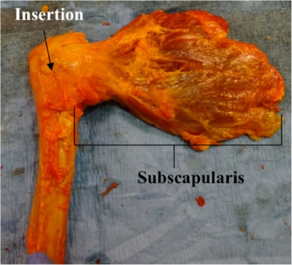
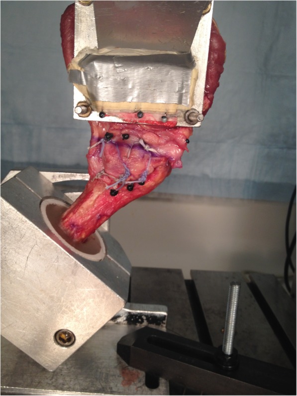
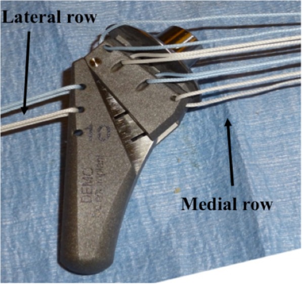
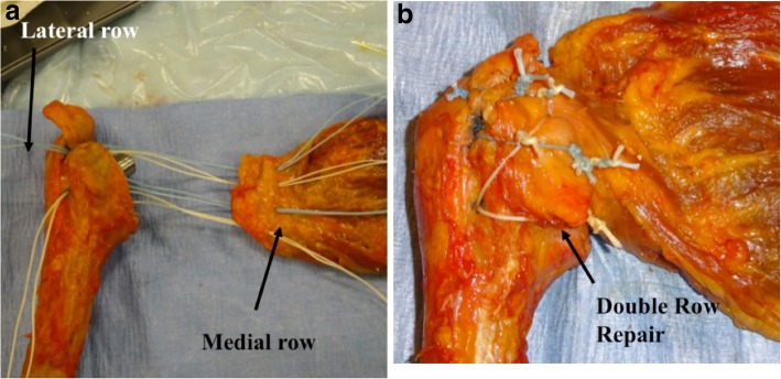
The subscapularis and proximal humerus were dissected from all soft tissues in 21 fresh-frozen human cadaveric shoulders and randomized to undergo SP, LTO, or standard subscapularis tenotomy (ST, control). For SP and LTO, six #5 sutures were passed through eyelets in the implant (on lateral border and through drill holes in bicipital groove [2] and under trunion [4]). Double-row repair was performed using two lateral row transosseous sutures and four medial row sutures through the tendon (SP) or osseotendinous junction (LTO). Biomechanical properties and mode of failure were tested.
There were no significant differences in elongation amplitude, cyclic elongation, or maximum load to failure between the three groups (P > 0.05). Mean stiffness was significantly higher in LTO (P = 0.009 vs. SP and ST). In the ST group, 7/7 specimens failed at the tendon-suture interface. For SP, 4/7 failed at the tendon-suture interface, one at the suture-bone interface, one fractured around the implant stem, and one at the knots. For LTO, 3/7 failed at the tendon-suture interface, two at the suture-bone interface and two fractured around the implant stem.
In this cadaveric model, subscapularis repair via ST, SP, and LTO techniques was biomechanically equivalent. Additional studies are needed to confirm these findings and determine the influence of biologic healing on healing rates and clinical outcomes.
N/a, biomechanical laboratory study.
在肩关节置换术中,肩胛下肌的处理存在争议。本研究的目的是比较肩胛下肌皮瓣(SP)和小结节截骨术(LTO)在尸体模型中的生物力学性能。
从 21 个新鲜冷冻的人体尸体肩关节的所有软组织中解剖出肩胛下肌和肱骨近端,并随机分为 SP、LTO 或标准肩胛下肌切断术(ST,对照组)。对于 SP 和 LTO,将六根 #5 缝线穿过植入物的眼孔(外侧缘和穿过二头肌沟的钻孔[2]和转子下[4])。采用双行外侧排贯穿骨缝线和四行内侧排缝线穿过肌腱(SP)或腱骨交界处(LTO)进行双排修复。测试生物力学性能和失效模式。
三组之间的伸长幅度、循环伸长和最大失效负荷无显著差异(P>0.05)。LTO 的平均刚度明显较高(P=0.009 与 SP 和 ST 相比)。在 ST 组中,7/7 个标本在肌腱-缝线界面处失效。对于 SP,有 4/7 个标本在肌腱-缝线界面处失效,1 个在缝线-骨界面处失效,1 个在植入物干周围断裂,1 个在结处失效。对于 LTO,有 3/7 个标本在肌腱-缝线界面处失效,2 个在缝线-骨界面处失效,2 个在植入物干周围断裂。
在这个尸体模型中,通过 ST、SP 和 LTO 技术进行的肩胛下肌修复在生物力学上是等效的。需要进一步的研究来证实这些发现,并确定生物愈合对愈合率和临床结果的影响。
无,生物力学实验室研究。