College of Animal Science and Veterinary Medicine, Jilin University, Changchun, China.
Laboratory of Biomolecular Research, Division of Biology and Chemistry, Paul Scherrer Institute, Villigen, Switzerland.
Front Immunol. 2019 Nov 14;10:2650. doi: 10.3389/fimmu.2019.02650. eCollection 2019.
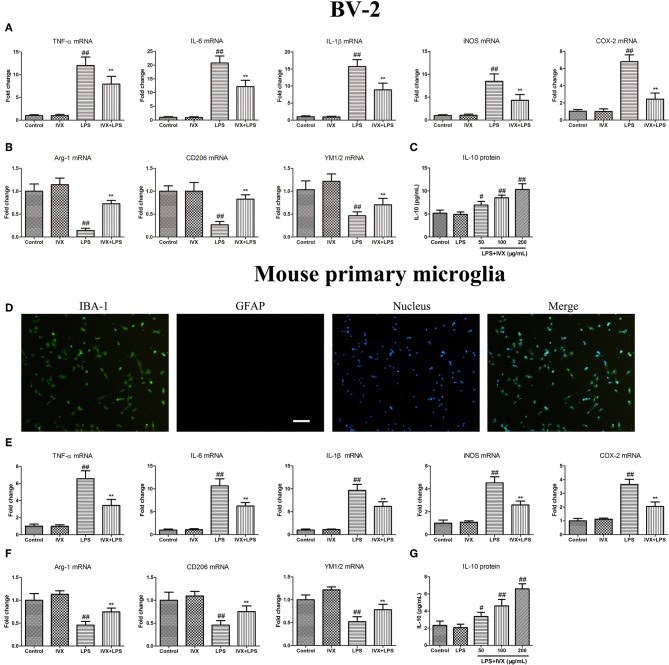
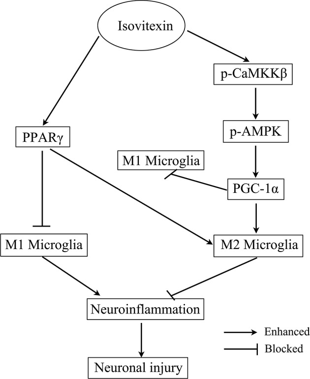
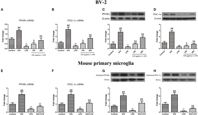
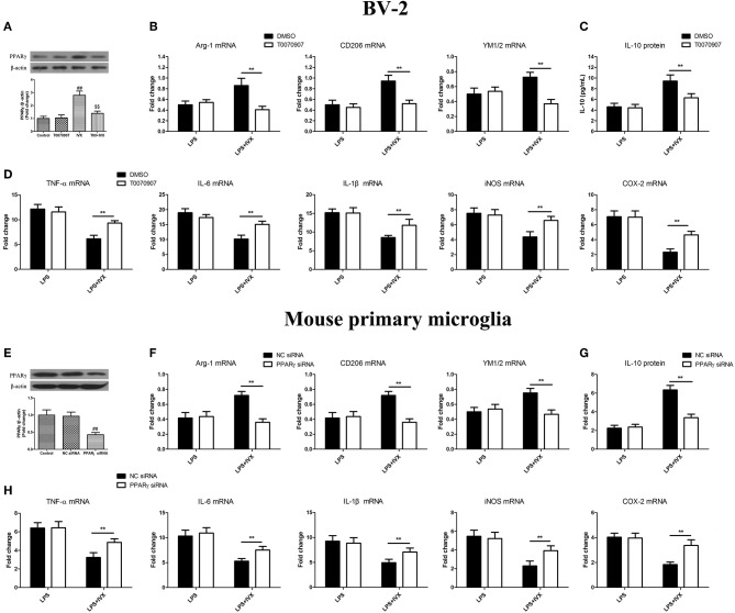
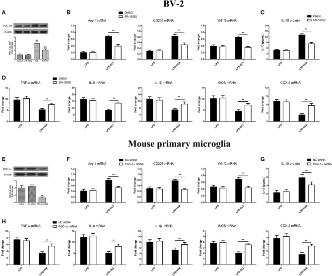
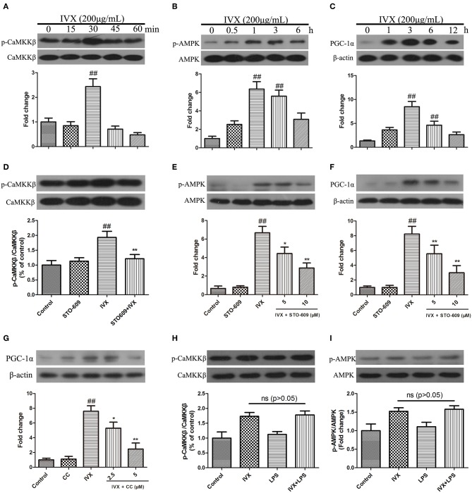
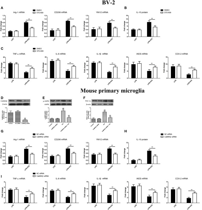
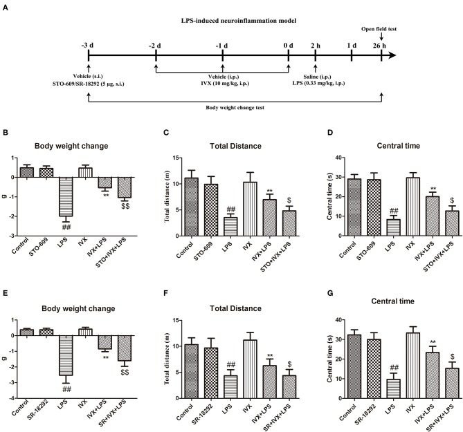
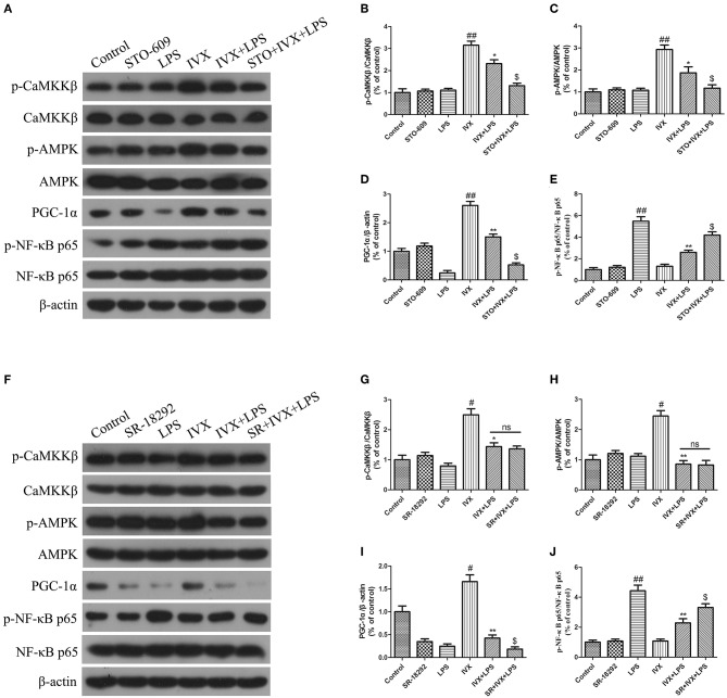
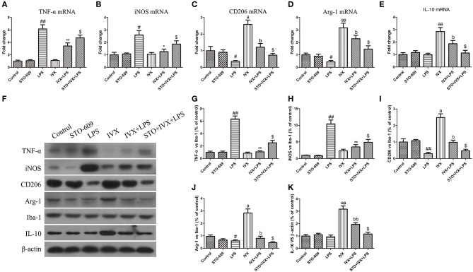
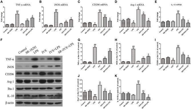
Microglia are the brain's immune cells and play an important role in regulating the microenvironment in the central nervous system. Activated microglia are capable of acquiring the pro-inflammatory (M1) phenotype and anti-inflammatory (M2) phenotype. Overactivation of microglia is neurotoxic and may lead to neuroinflammatory brain disorders. Neuroinflammation in the brain plays a crucial role part in the pathophysiology of many psychiatric and neurological diseases. The inhibition of M1 microglia and promotion of M2 microglia was demonstrated to treat and prevent these diseases through reduced neuroinflammation. Isovitexin (IVX) has anti-inflammatory properties and passes through the blood-brain barrier; however, the molecular mechanism that modulates IVX-mediated microglial polarization remains unclear. In BV-2 cells and mouse primary microglia, IVX suppressed the expression of M1 microglial markers, enhanced the expression of M2 microglial markers, and enhanced the release of interleukin 10 (IL-10). IVX promoted the expression of peroxisome proliferator-activated receptor-γ (PPARγ) and PPARγ coactivator-1α (PGC-1α) in LPS-induced microglial activation. The inhibition of PPARγ and PGC-1α attenuated the regulatory effect of IVX in LPS-induced microglial polarization. IVX increased the expression of p-CaMKKβ, p-AMPK, and PGC-1α in BV-2 cells. Inhibition of CaMKKβ with STO-609 or knockdown of CaMKKβ with CaMKKβ siRNA attenuated IVX-mediated M2 microglial polarization in LPS-treated cells. In LPS-treated mice, the inhibition of CaMKKβ and PGC-1α attenuated the IVX-mediated prevention of sickness behavior and enhanction of IVX-mediated M2 microglial polarization. IVX promoted M2 microglial polarization which exerted anti-inflammatory effects on LPS-induced neuroinflammation via the activation of the CaMKKβ/AMPK-PGC-1α signaling axis.
小胶质细胞是大脑的免疫细胞,在调节中枢神经系统的微环境中发挥重要作用。激活的小胶质细胞能够获得促炎(M1)表型和抗炎(M2)表型。小胶质细胞的过度激活具有神经毒性,可能导致神经炎症性脑疾病。大脑中的神经炎症在许多精神和神经疾病的病理生理学中起着至关重要的作用。通过减少神经炎症,抑制 M1 小胶质细胞和促进 M2 小胶质细胞被证明可以治疗和预防这些疾病。异牡荆素(IVX)具有抗炎特性并能穿过血脑屏障;然而,调节 IVX 介导的小胶质细胞极化的分子机制尚不清楚。在 BV-2 细胞和小鼠原代小胶质细胞中,IVX 抑制 M1 小胶质细胞标志物的表达,增强 M2 小胶质细胞标志物的表达,并增强白细胞介素 10(IL-10)的释放。IVX 在 LPS 诱导的小胶质细胞激活中促进过氧化物酶体增殖物激活受体-γ(PPARγ)和过氧化物酶体增殖物激活受体γ共激活物 1α(PGC-1α)的表达。PPARγ 和 PGC-1α 的抑制减弱了 IVX 在 LPS 诱导的小胶质细胞极化中的调节作用。IVX 增加了 BV-2 细胞中 p-CaMKKβ、p-AMPK 和 PGC-1α 的表达。用 STO-609 抑制 CaMKKβ 或用 CaMKKβ siRNA 敲低 CaMKKβ 可减弱 LPS 处理细胞中 IVX 介导的 M2 小胶质细胞极化。在 LPS 处理的小鼠中,抑制 CaMKKβ 和 PGC-1α 减弱了 IVX 介导的对疾病行为的预防和增强 IVX 介导的 M2 小胶质细胞极化。IVX 通过激活 CaMKKβ/AMPK-PGC-1α 信号通路促进 M2 小胶质细胞极化,从而对 LPS 诱导的神经炎症发挥抗炎作用。