Dipartimento di Scienze della Vita e Sanità Pubblica, Università Cattolica del Sacro Cuore, 00168 Rome, Italy.
Fondazione Policlinico Universitario A. Gemelli IRCCS, 00168 Rome, Italy.
Int J Mol Sci. 2019 Dec 25;21(1):175. doi: 10.3390/ijms21010175.
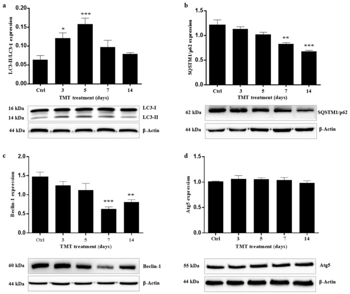
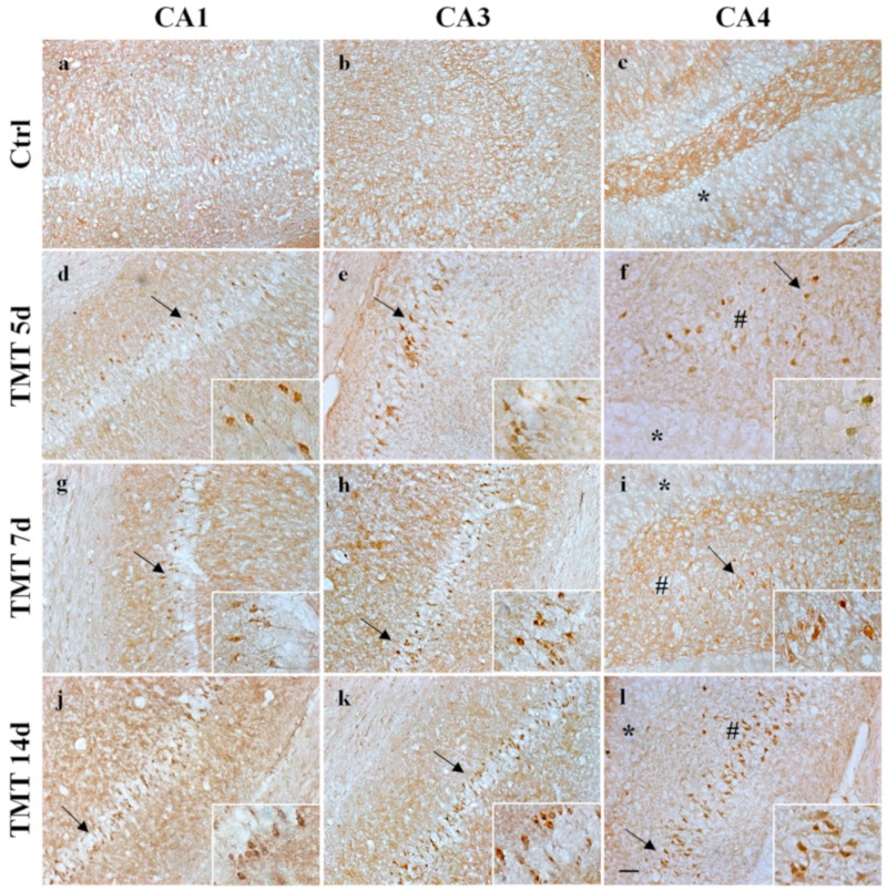
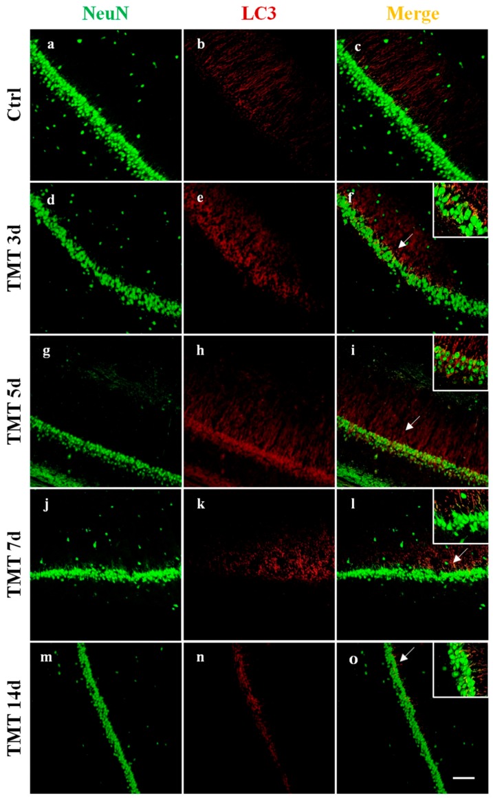
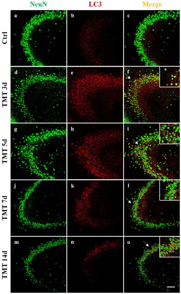
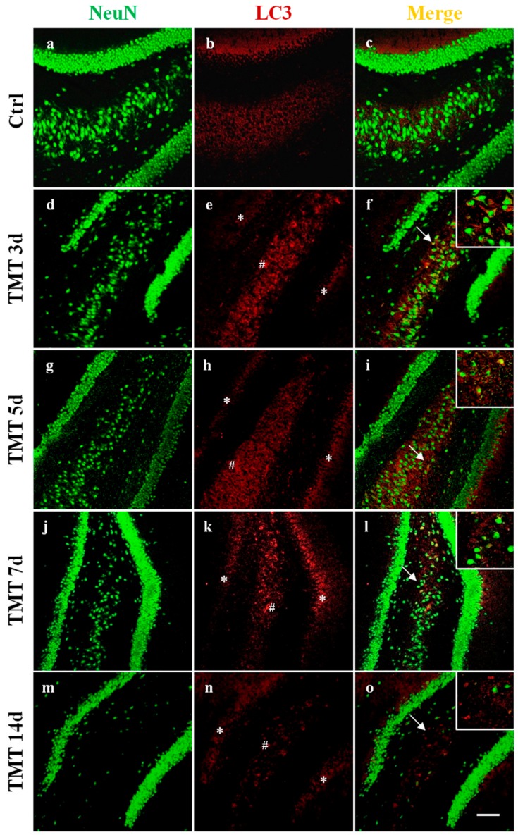
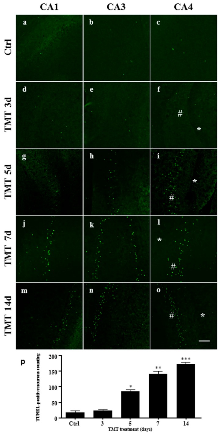
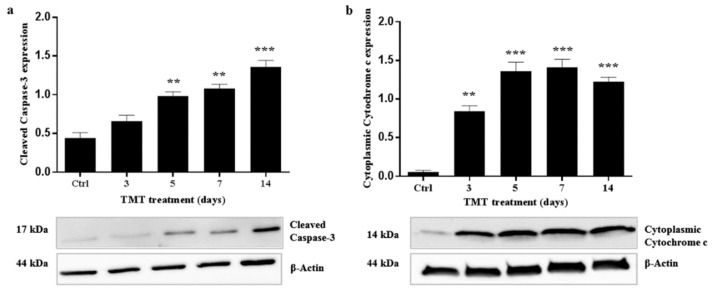
Trimethyltin (TMT) is an organotin compound known to produce significant and selective neuronal degeneration and reactive astrogliosis in the rodent central nervous system. Autophagy is the main cellular mechanism for degrading and recycling protein aggregates and damaged organelles, which in different stress conditions, such as starvation, generally improves cell survival. Autophagy is documented in several pathologic conditions, including neurodegenerative diseases. This study aimed to investigate the autophagy and apoptosis signaling pathways in hippocampal neurons of TMT-treated (Wistar) rats to explore molecular mechanisms involved in toxicant-induced neuronal injury. The microtubule-associated protein light chain (LC3, autophagosome marker) and sequestosome1 (SQSTM1/p62) (substrate of autophagy-mediated degradation) expressions were examined by Western blotting at different time points after intoxication. The results demonstrate that the LC3 II/I ratio significantly increased at 3 and 5 days, and that p62 levels significantly decreased at 7 and 14 days. Immunofluorescence images of LC3/neuronal nuclear antigen (NeuN) showed numerous strongly positive LC3 neurons throughout the hippocampus at 3 and 5 days. The terminal deoxynucleotidyltransferase dUTP nick end labeling (TUNEL) assay indicated an increase in apoptotic cells starting from 5 days after treatment. In order to clarify apoptotic pathway, immunofluorescence images of apoptosis-inducing factor (AIF)/NeuN did not show nuclear translocation of AIF in neurons. Increased expression of cleaved Caspase-3 was revealed at 5-14 days in all hippocampal regions by Western blotting and immunohistochemistry analyses. These data clearly demonstrate that TMT intoxication induces a marked increase in both autophagy and caspase-dependent apoptosis, and that autophagy occurring just before apoptosis could have a potential role in neuronal loss in this experimental model of neurodegeneration.
三甲基锡(TMT)是一种有机锡化合物,已知它会导致啮齿动物中枢神经系统中出现显著的、选择性的神经元变性和反应性星形胶质增生。自噬是降解和回收蛋白质聚集体和受损细胞器的主要细胞机制,在饥饿等不同应激条件下,通常会提高细胞存活率。自噬在几种病理条件下都有记录,包括神经退行性疾病。本研究旨在研究 TMT 处理(Wistar)大鼠海马神经元中的自噬和细胞凋亡信号通路,以探索参与毒物诱导的神经元损伤的分子机制。在中毒后不同时间点通过 Western blot 检测微管相关蛋白轻链(LC3,自噬体标志物)和自噬体底物 1(SQSTM1/p62)的表达。结果表明,LC3 II/I 比值在 3 天和 5 天显著增加,p62 水平在 7 天和 14 天显著降低。LC3/神经元核抗原(NeuN)的免疫荧光图像显示,在 3 天和 5 天,整个海马区有大量强烈阳性的 LC3 神经元。末端脱氧核苷酸转移酶 dUTP 缺口末端标记(TUNEL)检测表明,从治疗后 5 天开始,凋亡细胞数量增加。为了阐明细胞凋亡途径,凋亡诱导因子(AIF)/NeuN 的免疫荧光图像显示神经元中 AIF 没有发生核转位。Western blot 和免疫组织化学分析显示,在所有海马区,Cleaved Caspase-3 的表达在 5-14 天增加。这些数据清楚地表明,TMT 中毒会导致自噬和 caspase 依赖性细胞凋亡明显增加,并且在这个神经退行性变的实验模型中,凋亡前发生的自噬可能在神经元丢失中发挥潜在作用。