Liu Jingjing, Shi Yupeng, Han Jing, Zhang Yong, Cao Zhenghao, Cheng Jingliang
Department of MRI, The First Affiliated Hospital of Zhengzhou University, Zhengzhou, China.
Department of Pathology, The First Affiliated Hospital of Zhengzhou University, Zhengzhou, China.
Int J Stem Cells. 2020 Mar 30;13(1):104-115. doi: 10.15283/ijsc19098.
Tracking of the tumor progression by MSCs-based therapy is being increasingly important in evaluating relative therapy effectively. Herein, Bioluminescence imaging (BLI) technology was used to dynamically and quantitatively track the hepatocellular carcinoma suppressive effects by human umbilical cord mesenchymal stem cells (UC-MSCs).
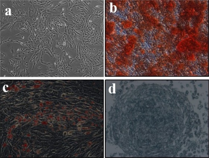
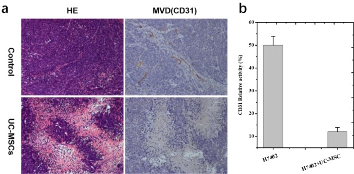
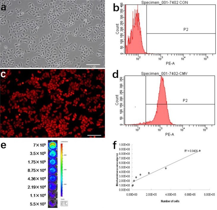
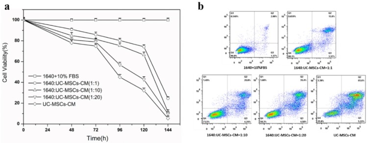
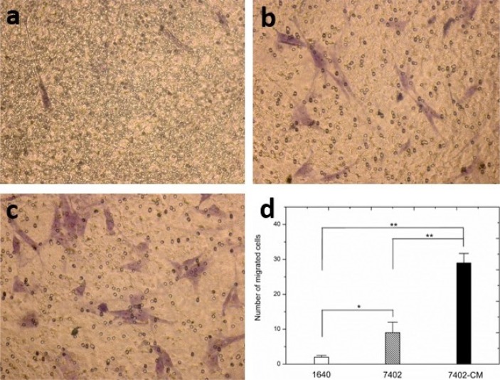
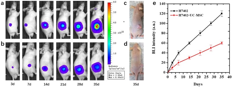
The stem cells present typical phenotypic characteristics and differentiation ability by morphology and flow cytometry analysis of marker expression. Then, the growth inhibition effect of conditioned medium and UC-MSC on H7402 cells was studied. It is found both the conditioned medium and UC-MSC can effectively decrease the proliferation of H7402 cells compared with the control group. Meanwhile, the relative migration of UC-MSC to H7402 is also increased through the transwell migration assay. In addition, a mice hepatoma tumor model was built by H7402 cells which can express a pLenti-6.3/DEST-CMV-luciferase 2-mKate2 gene. The effect of stem cells on growth inhibition of tumor in a mice transplantation model was dynamically monitored by bioluminescence imaging within 5 weeks. It has shown the bioluminescence signal intensity of the tumor model was significantly higher than that of the UC-MSC co-acting tumor model, indicating that the inhibition of UC-MSC on liver cancer resulted in low expression of bioluminescent signals.
The microenvironment of UC-MSCs can effectively inhibit the growth of liver cancer cells, and this therapeutic effect can be dynamically and quantitatively monitored in vivo by BLI. This is of great significance for the imaging research and application of stem cells in anticancer therapy.
基于间充质干细胞(MSCs)的治疗对肿瘤进展的追踪在有效评估相关治疗中变得越来越重要。在此,利用生物发光成像(BLI)技术动态定量追踪人脐带间充质干细胞(UC-MSCs)对肝癌的抑制作用。
通过形态学及标记物表达的流式细胞术分析,干细胞呈现出典型的表型特征和分化能力。然后,研究了条件培养基和UC-MSC对H7402细胞的生长抑制作用。发现与对照组相比,条件培养基和UC-MSC均能有效降低H7402细胞的增殖。同时,通过Transwell迁移实验也发现UC-MSC向H7402的相对迁移增加。此外,用可表达pLenti-6.3/DEST-CMV-荧光素酶2-mKate2基因的H7402细胞建立小鼠肝癌肿瘤模型。在5周内通过生物发光成像动态监测干细胞对小鼠移植模型中肿瘤生长抑制的作用。结果显示肿瘤模型的生物发光信号强度明显高于UC-MSC共同作用的肿瘤模型,表明UC-MSC对肝癌的抑制导致生物发光信号低表达。
UC-MSCs的微环境能有效抑制肝癌细胞生长,且这种治疗效果可通过BLI在体内进行动态定量监测。这对干细胞在抗癌治疗中的成像研究及应用具有重要意义。