Xu Meifang, Wang Fei, Chen Hong, Liu Lin, Liu Wenwen, Yang Yinghong, Zheng Qiaoling, Zhang Lihong, Li Xiaoxuan, Lin Suxia, Zang Shengbing
Department of Pathology, Fujian Medical University Union Hospital, Fuzhou 350001, China.
Division of Pediatrics, University of Texas MD Anderson Cancer Center, Houston, TX 77030, USA.
Cancer Biol Med. 2019 Nov;16(4):743-755. doi: 10.20892/j.issn.2095-3941.2019.0115.
Angioimmunoblastic T cell lymphoma (AITL) is an aggressive form of non-Hodgkin lymphoma derived from mature T cells. However, the underlying pathogenesis of AITL remains unresolved. We aimed to explore the role of FOXO1-mediated signaling in the tumorigenesis and progression of AITL.
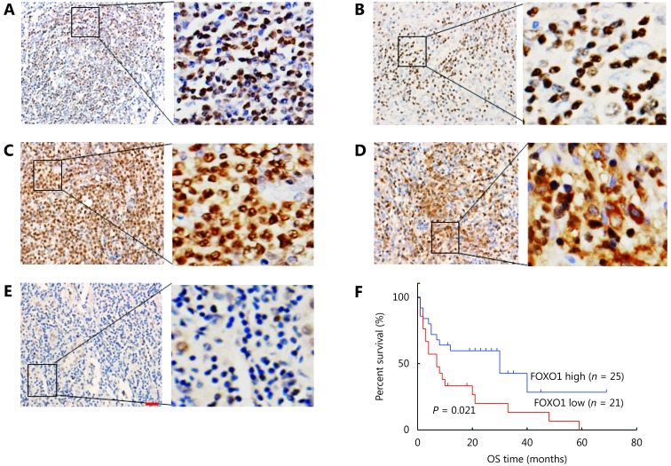
FOXO1 expression was assessed using immunohistochemistry on a total of 46 AITL tissue samples. Retroviruses encoding FOXO1 shRNA were used to knockdown FOXO1 expression in CD4 T cells. Flow cytometric assays analyzed the proliferation and survival of FOXO1 knockdown CD4 T cells. Furthermore, we performed adoptive T-cell transfer experiments to identify whether inactivation of FOXO1 induced neoplastic follicular-helper T (Tfh) cell polarization and function.
Patients with low FOXO1 protein levels were prone to have an advanced tumor stage ( = 0.049), higher ECOG ps ( = 0.024), the presence of bone marrow invasion ( = 0.000), and higher IPI ( = 0.035). Additionally, the survival rates of patients in the FOXO1 high-expression group were significantly better than those in the FOXO1 low-expression group ( = 5.346, = 0.021). We also observed that inactivation of FOXO1 increased CD4 T cell proliferation and altered the survival and cell-cycle progression of CD4 T cells. Finally, we confirmed that inactivation of FOXO1 induces Tfh cell programing and function.
Inactivation of FOXO1 in AITL plays a key role in the tumorigenesis and progression of AITL. We propose that FOXO1 expression could be a useful prognostic marker in AITL patients to predict poor survival, and to design appropriate therapeutic strategies.
血管免疫母细胞性T细胞淋巴瘤(AITL)是一种源自成熟T细胞的侵袭性非霍奇金淋巴瘤。然而,AITL的潜在发病机制仍未明确。我们旨在探讨FOXO1介导的信号通路在AITL肿瘤发生和进展中的作用。
采用免疫组织化学方法对46例AITL组织样本进行FOXO1表达评估。使用编码FOXO1短发夹RNA的逆转录病毒敲低CD4 T细胞中的FOXO1表达。流式细胞术分析FOXO1敲低的CD4 T细胞的增殖和存活情况。此外,我们进行了过继性T细胞转移实验,以确定FOXO1失活是否诱导肿瘤滤泡辅助性T(Tfh)细胞极化和功能。
FOXO1蛋白水平低的患者更容易出现肿瘤晚期(P = 0.049)、较高的东部肿瘤协作组体能状态(P = 0.024)、骨髓侵犯(P = 0.000)以及较高的国际预后指数(P = 0.035)。此外,FOXO1高表达组患者的生存率明显优于FOXO1低表达组(P = 5.346,P = 0.021)。我们还观察到FOXO1失活增加了CD4 T细胞增殖,并改变了CD4 T细胞的存活和细胞周期进程。最后,我们证实FOXO1失活诱导Tfh细胞编程和功能。
AITL中FOXO1失活在AITL的肿瘤发生和进展中起关键作用。我们提出FOXO1表达可能是AITL患者预测不良生存和设计合适治疗策略的有用预后标志物。