Centre for Operative Dentistry, Periodontology, and Endodontology, University of Dental Medicine and Oral Health, Danube Private University, Krems, Austria.
Department for Biomedical Research, Danube University, Krems, Austria.
PLoS One. 2020 Jan 28;15(1):e0228249. doi: 10.1371/journal.pone.0228249. eCollection 2020.
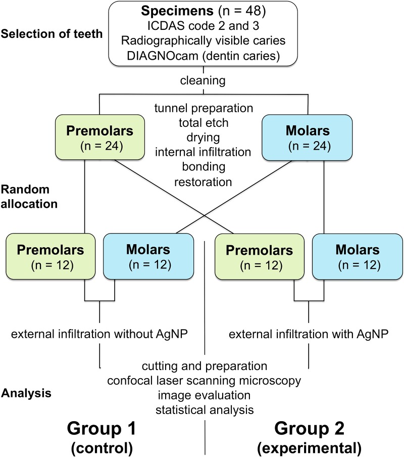
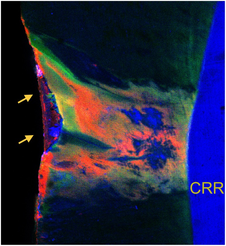
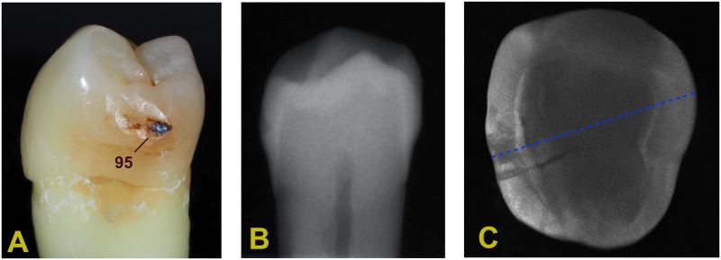
This ex vivo proof-of-concept study aimed to investigate the effect of nanosilver particles (AgNP) added to a conventional infiltrant resin (Icon) on external penetration into natural proximal enamel caries exceeding into dentin after internal tunnel preparation and internal infiltration. Carious lesions (ICDAS codes 2/3) of extracted human (pre-)molars revealing proximal caries radiographically exceeding into dentin (E2/D1 lesions) were preselected. Then, 48 of those specimens showing demineralized areas transcending the enamel-dentin border as assessed by means of near-infrared light transillumination (DIAGNOcam) were deproteinized (NaOCl, 5%). Using an internal tunnel approach, occlusal cavities central to the marginal ridge were prepared. Excavation of carious dentin, total etch procedure (H3PO4, 40%), and internal resin infiltration (FITC-labeled) followed, along with final restorations (flowable composite resin). Outer lesion surfaces were etched (HCl, 15%) prior to external infiltration (RITC-labeled). Group 1 (control; n = 24) used non-modified infiltrant, while an infiltrant/AgNP mixture (20 nm; 5.5 wt%) was used with experimental Group 2 (n = 24). Non-infiltrated pores of cut lesions were stained (Berberine), and specimens were analyzed using confocal laser scanning microscopy. Compared to the non-filled infiltrant, incorporation of AgNP had no effect on the resin's external penetration. Between the groups, no significant differences regarding internal or external infiltration could be detected, and non-infiltrated lesion areas did not differ significantly (p>0.109; t-test). The internal tunnel preparation in combination with both an internal resin infiltration and an additional external infiltration approach using a nanosilver-modified infiltrant resin leads to increased infiltrated lesion areas, thus occluding and adhesively stabilizing the porous volume of the demineralized enamel. While exerting antimicrobial effects by the nanosilver particles, this approach should have the potential as a viable treatment alternative for proximal lesions extending into dentin, thus avoiding the sacrifice of sound enamel, postponing the frequently inevitable restoration/re-restoration cycle of conventional proximal caries treatment, and improving dental health.
本离体概念验证研究旨在探讨在内部隧道制备和内部渗透后,添加纳米银颗粒 (AgNP) 对常规渗透树脂 (Icon) 进入天然近釉质龋的外部渗透的影响,这些龋超过牙本质。从离体的(前)磨牙中预先选择显示出放射状超过牙本质的近釉质龋(E2/D1 病变)的龋损(ICDAS 代码 2/3)。然后,使用近红外光透射(DIAGNOcam)评估,选择 48 个显示出脱矿区域超过釉牙本质边界的标本进行去蛋白(NaOCl,5%)。使用内部隧道方法,在牙尖边缘中央制备窝洞。然后进行龋齿的挖除、全酸蚀处理(H3PO4,40%)和内部树脂渗透(FITC 标记),以及最终的修复(流动复合树脂)。在外部渗透(RITC 标记)之前,对外部病变表面进行酸蚀(HCl,15%)。第 1 组(对照组;n = 24)使用未改性的渗透剂,而第 2 组(实验组;n = 24)使用渗透剂/AgNP 混合物(20nm;5.5wt%)。用盐酸(15%)酸蚀切割病变的非渗透孔,并使用共聚焦激光扫描显微镜对标本进行分析。与未填充的渗透剂相比,AgNP 的掺入对树脂的外部渗透没有影响。在两组之间,对内、外渗透都没有发现显著差异,非渗透病变区域也没有显著差异(p>0.109;t 检验)。内部隧道制备结合内部树脂渗透和使用纳米银改性渗透树脂的额外外部渗透方法,增加了渗透病变区域,从而封闭和粘合稳定脱矿釉质的多孔体积。虽然纳米银颗粒发挥了抗菌作用,但这种方法作为一种可行的替代治疗方法,适用于扩展到牙本质的近釉质龋,从而避免牺牲健康的釉质,推迟传统近釉质龋治疗中经常不可避免的修复/再修复周期,并改善口腔健康。