Pan Pan, Wei Shubao, Ou Yangpan, Jiang Wenyan, Li Wenmei, Lei Yiwu, Liu Feng, Guo Wenbin, Luo Shuguang
Department of Psychiatry, The Second Xiangya Hospital of Central South University, Changsha, China.
National Clinical Research Center on Mental Disorders, Changsha, China.
Front Neurol. 2020 Jan 10;10:1358. doi: 10.3389/fneur.2019.01358. eCollection 2019.
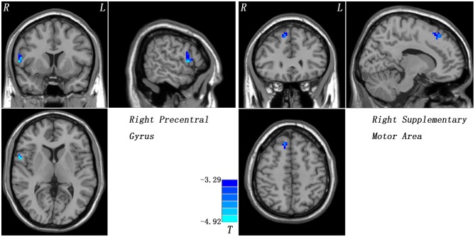
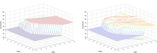
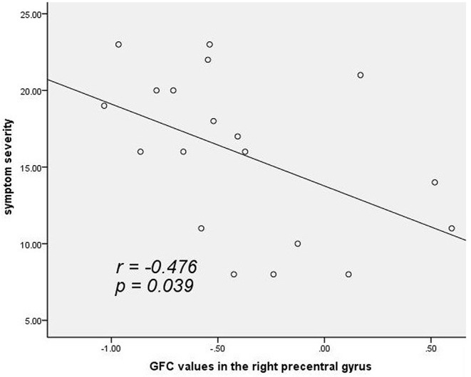
Altered functional connectivity (FC) is related to pathophysiology of patients with cervical dystonia (CD). However, inconsistent results may be obtained due to different selected regions of interest. We explored voxel-wise brain-wide FC changes in patients with CD at rest in an unbiased manner and analyzed their correlations with symptomatic severity using the Tsui scale. A total of 19 patients with CD and 21 sex- and age-matched healthy controls underwent resting-state functional magnetic resonance imaging scans. Global-brain FC (GFC) was applied to analyze the images. Support vector machine was used to distinguish the patients from the controls. Patients with CD exhibited decreased GFC in the right precentral gyrus and right supplementary motor area (SMA) that belonged to the M1-SMA motor network. Significantly negative correlation was observed between GFC values in the right precentral gyrus and symptomatic severity in the patients ( = -0.476, = 0.039, uncorrected). Decreased GFC values in these two brain regions could be utilized to differentiate the patients from the controls with good accuracies, sensitivities and specificities (83.33, 85.71, and 80.95% in the right precentral gyrus; and 87.59, 89.49, and 85.71% in the right SMA). Our investigation suggests that patients with CD show reduced GFC in brain regions of the M1-SMA motor network and provides further insights into the pathophysiology of CD. GFC values in the right precentral gyrus and right SMA may be used as potential biomarkers to recognize the patients from the controls.
功能连接性(FC)改变与颈部肌张力障碍(CD)患者的病理生理学相关。然而,由于感兴趣区域选择不同,可能会得到不一致的结果。我们以无偏倚的方式探索了CD患者静息状态下全脑体素水平的FC变化,并使用徐氏量表分析其与症状严重程度的相关性。共有19例CD患者和21例性别及年龄匹配的健康对照者接受了静息态功能磁共振成像扫描。采用全脑功能连接性(GFC)分析图像。使用支持向量机区分患者和对照者。CD患者在属于M1-SMA运动网络的右侧中央前回和右侧辅助运动区(SMA)表现出GFC降低。在患者中观察到右侧中央前回的GFC值与症状严重程度之间存在显著负相关(r = -0.476,P = 0.039,未校正)。这两个脑区GFC值降低可用于以良好的准确性、敏感性和特异性区分患者和对照者(右侧中央前回分别为83.33%、85.71%和80.95%;右侧SMA分别为87.59%、89.49%和85.71%)。我们的研究表明,CD患者在M1-SMA运动网络的脑区表现出GFC降低,并为CD的病理生理学提供了进一步的见解。右侧中央前回和右侧SMA的GFC值可能用作区分患者和对照者的潜在生物标志物。