• 文献检索
  • 文档翻译
  • 深度研究
  • 学术资讯
  • Suppr Zotero 插件Zotero 插件
  • 邀请有礼
  • 套餐&价格
  • 历史记录
应用&插件
Suppr Zotero 插件Zotero 插件浏览器插件Mac 客户端Windows 客户端微信小程序
定价
高级版会员购买积分包购买API积分包
服务
文献检索文档翻译深度研究API 文档MCP 服务
关于我们
关于 Suppr公司介绍联系我们用户协议隐私条款
关注我们

Suppr 超能文献

核心技术专利:CN118964589B侵权必究
粤ICP备2023148730 号-1Suppr @ 2026

文献检索

告别复杂PubMed语法,用中文像聊天一样搜索,搜遍4000万医学文献。AI智能推荐,让科研检索更轻松。

立即免费搜索

文件翻译

保留排版,准确专业,支持PDF/Word/PPT等文件格式,支持 12+语言互译。

免费翻译文档

深度研究

AI帮你快速写综述,25分钟生成高质量综述,智能提取关键信息,辅助科研写作。

立即免费体验

在低度全身性炎症期间,单核细胞介导循环微泡归巢至肺血管系统。

Monocytes mediate homing of circulating microvesicles to the pulmonary vasculature during low-grade systemic inflammation.

作者信息

O'Dea Kieran P, Tan Ying Ying, Shah Sneh, V Patel Brijesh, C Tatham Kate, Wilson Mike R, Soni Sanooj, Takata Masao

机构信息

Section of Anaesthetics, Pain Medicine & Intensive Care, Imperial College London, Chelsea & Westminster Hospital, London, UK.

出版信息

J Extracell Vesicles. 2020 Jan 5;9(1):1706708. doi: 10.1080/20013078.2019.1706708. eCollection 2020.

DOI:10.1080/20013078.2019.1706708
PMID:32002170
原文链接:https://pmc.ncbi.nlm.nih.gov/articles/PMC6968433/
Abstract

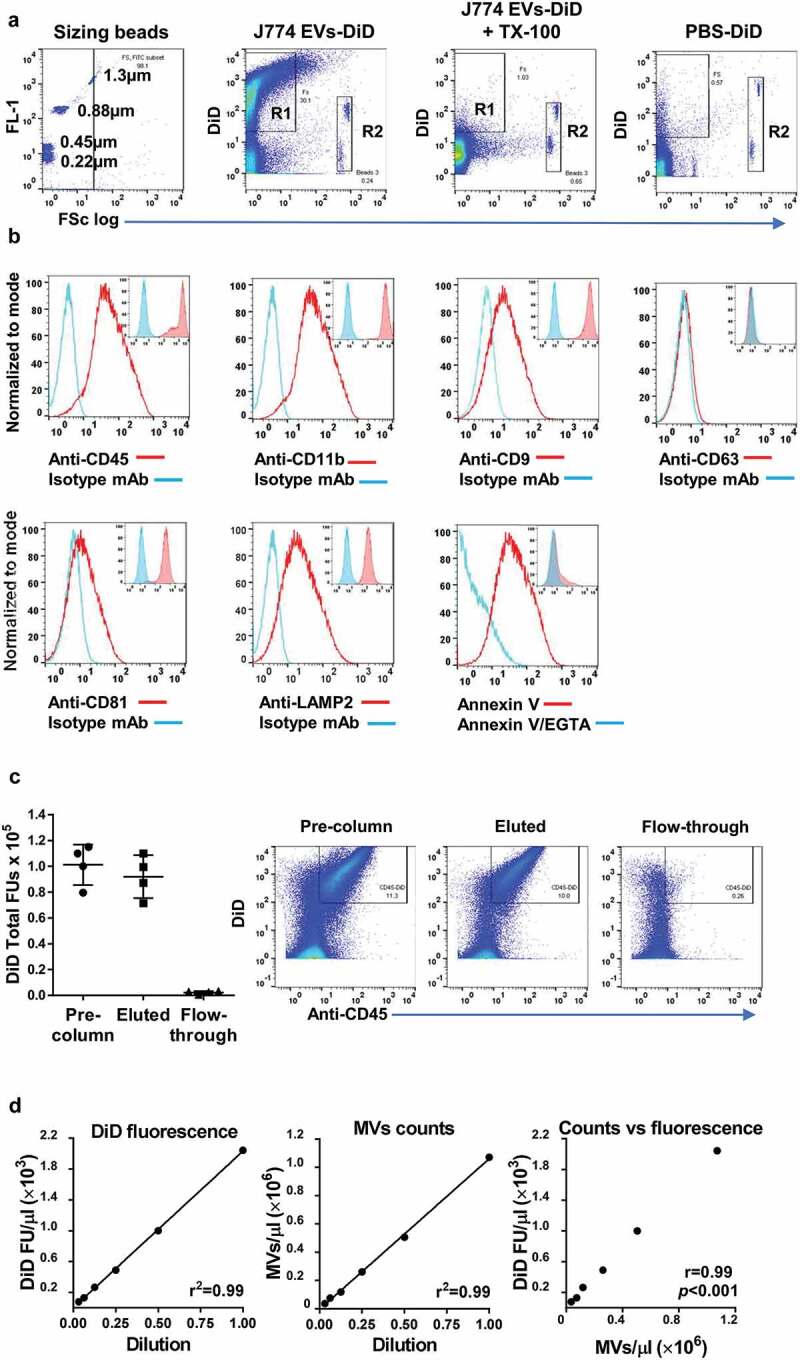
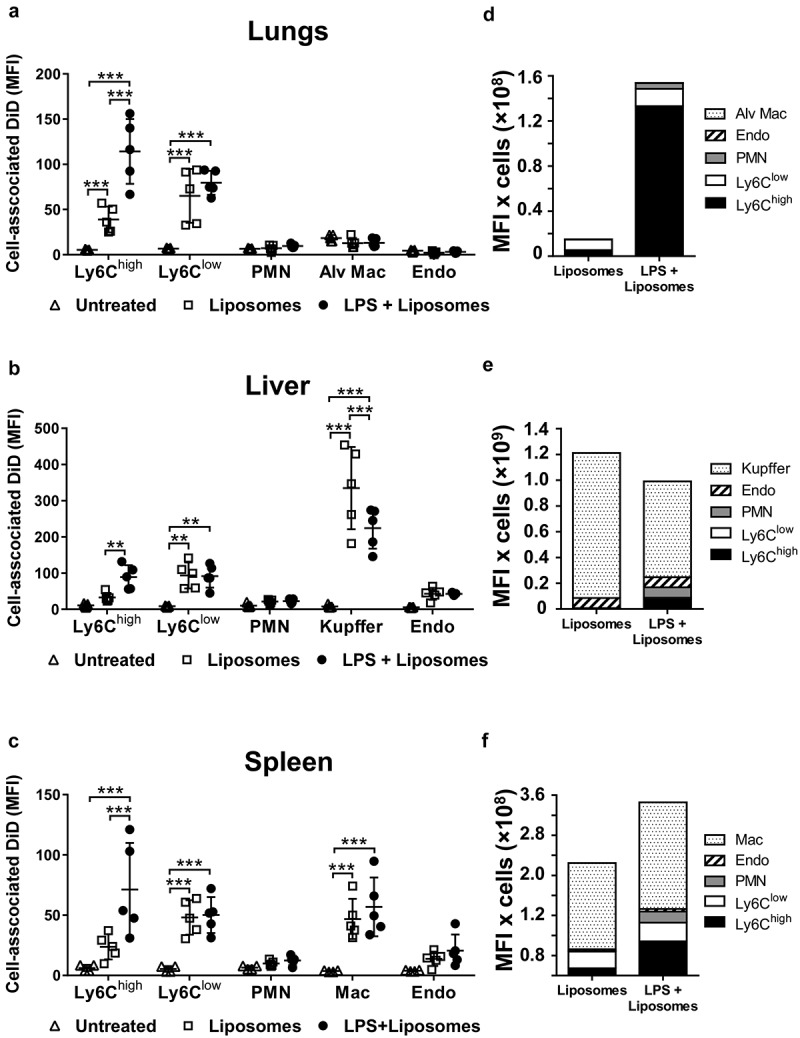
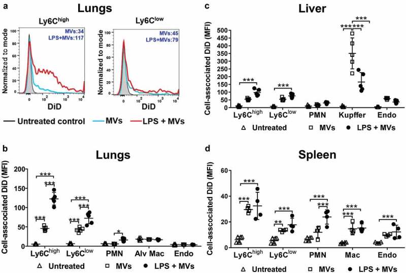
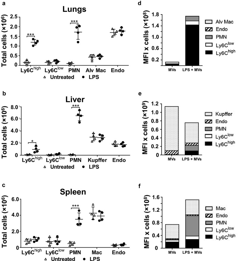
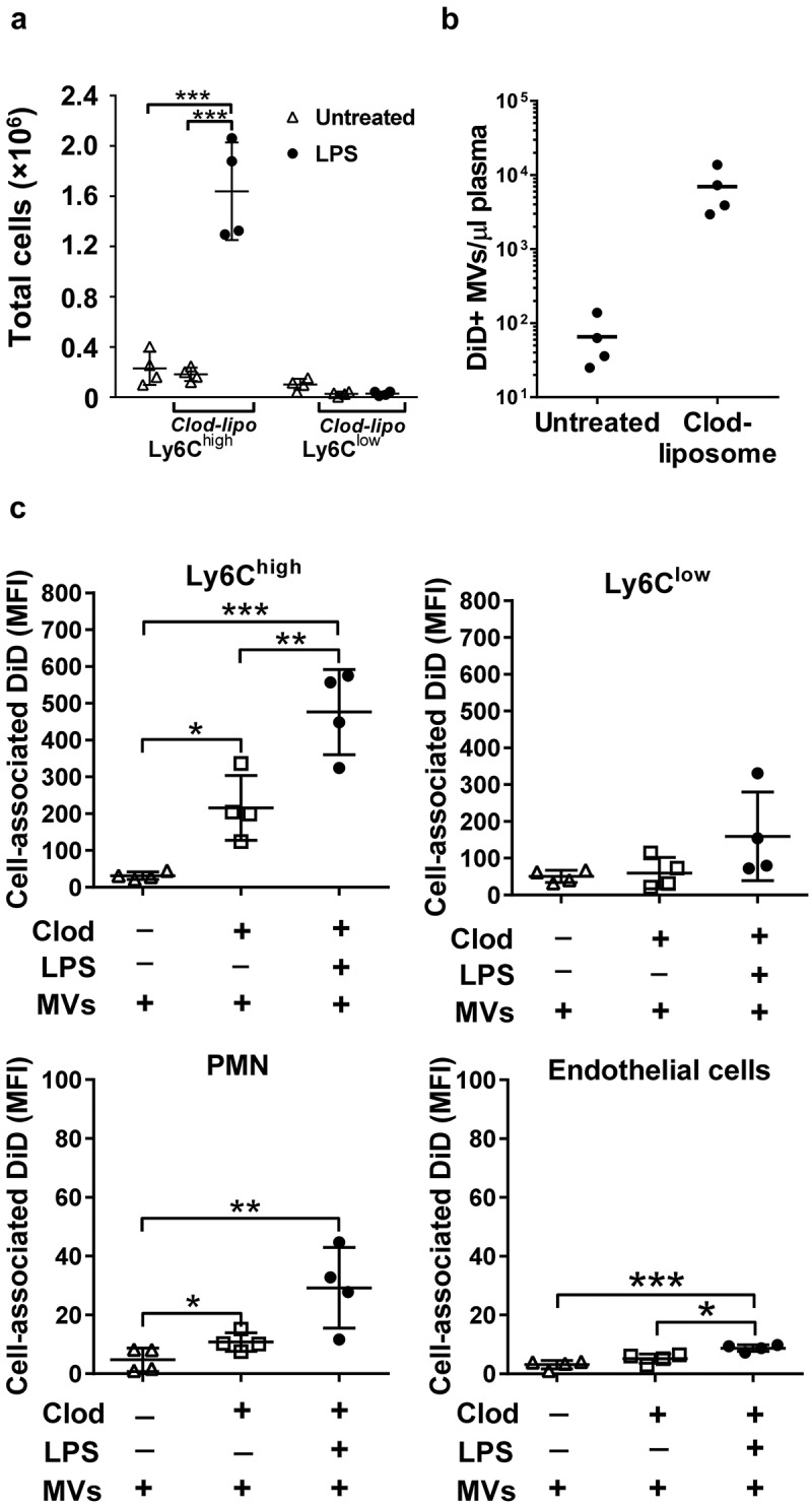
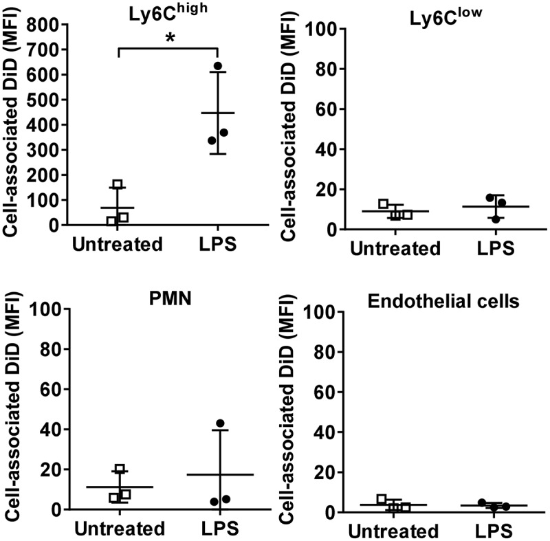
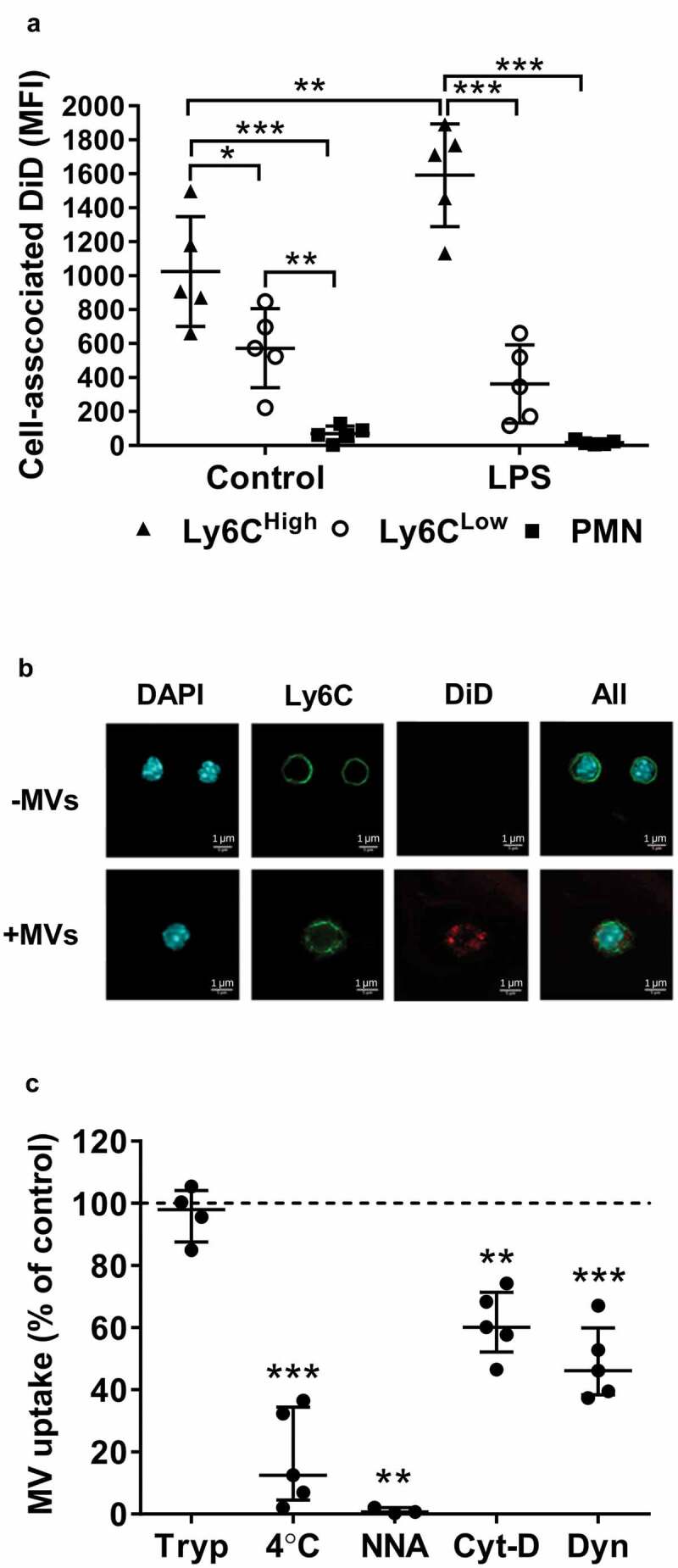
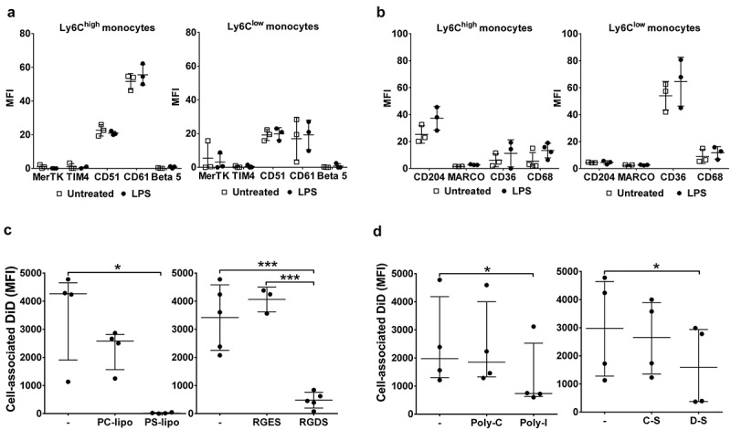
Microvesicles (MVs), a plasma membrane-derived subclass of extracellular vesicles, are produced and released into the circulation during systemic inflammation, yet little is known of cell/tissue-specific uptake of MVs under these conditions. We hypothesized that monocytes contribute to uptake of circulating MVs and that their increased margination to the pulmonary circulation and functional priming during systemic inflammation produces substantive changes to the systemic MV homing profile. Cellular uptake of i.v.-injected, fluorescently labelled MVs (J774.1 macrophage-derived) in vivo was quantified by flow cytometry in vascular cell populations of the lungs, liver and spleen of C57BL6 mice. Under normal conditions, both Ly6C and Ly6C monocytes contributed to MV uptake but liver Kupffer cells were the dominant target cell population. Following induction of sub-clinical endotoxemia with low-dose i.v. LPS, MV uptake by lung-marginated Ly6C monocytes increased markedly, both at the individual cell level (2.5-fold) and through substantive expansion of their numbers (8-fold), whereas uptake by splenic macrophages was unchanged and uptake by Kupffer cells actually decreased (~50%). Further analysis of MV uptake within the pulmonary vasculature using a combined model approach of in vivo macrophage depletion, ex vivo isolated perfused lungs and in vitro lung perfusate cell-based assays, indicated that Ly6C monocytes possess a high MV uptake capacity (equivalent to Kupffer cells), that is enhanced directly by endotoxemia and ablated in the presence of phosphatidylserine (PS)-enriched liposomes and β3 integrin receptor blocking peptide. Accordingly, i.v.-injected PS-enriched liposomes underwent a redistribution of cellular uptake during endotoxemia similar to MVs, with enhanced uptake by Ly6C monocytes and reduced uptake by Kupffer cells. These findings indicate that monocytes, particularly lung-marginated Ly6C subset monocytes, become a dominant target cell population for MVs during systemic inflammation, with significant implications for the function and targeting of endogenous and therapeutically administered MVs, lending novel insights into the pathophysiology of pulmonary vascular inflammation.

摘要

微泡(MVs)是细胞外囊泡的一个源自质膜的亚类,在全身炎症期间产生并释放到循环中,但在这些情况下,关于MVs的细胞/组织特异性摄取知之甚少。我们假设单核细胞有助于循环MVs的摄取,并且它们在全身炎症期间增加向肺循环的边缘化和功能启动会导致全身MV归巢谱发生实质性变化。通过流式细胞术对C57BL6小鼠肺、肝和脾的血管细胞群体中静脉注射的荧光标记MVs(J774.1巨噬细胞来源)的细胞摄取进行定量。在正常条件下,Ly6C单核细胞和Ly6C⁻单核细胞都有助于MV摄取,但肝库普弗细胞是主要的靶细胞群体。在用低剂量静脉注射脂多糖诱导亚临床内毒素血症后,肺边缘的Ly6C单核细胞对MV的摄取在个体细胞水平上显著增加(约2.5倍),并且通过其数量的实质性增加(约8倍),而脾巨噬细胞的摄取没有变化,库普弗细胞的摄取实际上减少了(约50%)。使用体内巨噬细胞耗竭、离体分离灌注肺和基于体外肺灌注液细胞的检测的联合模型方法对肺血管内MV摄取的进一步分析表明,Ly6C单核细胞具有高MV摄取能力(等同于库普弗细胞),内毒素血症可直接增强该能力,并且在存在富含磷脂酰丝氨酸(PS)的脂质体和β₃整合素受体阻断肽的情况下被消除。因此,静脉注射的富含PS的脂质体在内毒素血症期间经历了与MVs类似的细胞摄取重新分布,Ly6C单核细胞的摄取增加,库普弗细胞的摄取减少。这些发现表明,单核细胞,特别是肺边缘的Ly6C亚群单核细胞,在全身炎症期间成为MVs的主要靶细胞群体,这对内源性和治疗性施用的MVs的功能和靶向具有重要意义,为肺血管炎症的病理生理学提供了新的见解。

https://cdn.ncbi.nlm.nih.gov/pmc/blobs/5296/6968433/c2ac260c935b/ZJEV_A_1706708_F0008_B.jpg
https://cdn.ncbi.nlm.nih.gov/pmc/blobs/5296/6968433/e4d05147f406/ZJEV_A_1706708_F0001_OC.jpg
https://cdn.ncbi.nlm.nih.gov/pmc/blobs/5296/6968433/803ba677769e/ZJEV_A_1706708_F0002_OC.jpg
https://cdn.ncbi.nlm.nih.gov/pmc/blobs/5296/6968433/5038d5cdf7f7/ZJEV_A_1706708_F0003_B.jpg
https://cdn.ncbi.nlm.nih.gov/pmc/blobs/5296/6968433/820ef0a7c410/ZJEV_A_1706708_F0004_B.jpg
https://cdn.ncbi.nlm.nih.gov/pmc/blobs/5296/6968433/43fecd6cd1c6/ZJEV_A_1706708_F0005_B.jpg
https://cdn.ncbi.nlm.nih.gov/pmc/blobs/5296/6968433/2b094db9662c/ZJEV_A_1706708_F0006_OC.jpg
https://cdn.ncbi.nlm.nih.gov/pmc/blobs/5296/6968433/c951ce3d3ba1/ZJEV_A_1706708_F0007_B.jpg
https://cdn.ncbi.nlm.nih.gov/pmc/blobs/5296/6968433/c2ac260c935b/ZJEV_A_1706708_F0008_B.jpg

相似文献

1
Monocytes mediate homing of circulating microvesicles to the pulmonary vasculature during low-grade systemic inflammation.在低度全身性炎症期间,单核细胞介导循环微泡归巢至肺血管系统。
J Extracell Vesicles. 2020 Jan 5;9(1):1706708. doi: 10.1080/20013078.2019.1706708. eCollection 2020.
2
Investigation of Microvesicle Uptake by Mouse Lung-marginated Monocytes .小鼠肺边缘单核细胞对微泡摄取的研究
Bio Protoc. 2022 Feb 5;12(3):e4307. doi: 10.21769/BioProtoc.4307.
3
Microvesicle-Mediated Communication Within the Alveolar Space: Mechanisms of Uptake by Epithelial Cells and Alveolar Macrophages.肺泡腔内的微小囊泡介导的通讯:上皮细胞和肺泡巨噬细胞摄取的机制。
Front Immunol. 2022 Apr 27;13:853769. doi: 10.3389/fimmu.2022.853769. eCollection 2022.
4
Cholesterol enrichment of human monocyte/macrophages induces surface exposure of phosphatidylserine and the release of biologically-active tissue factor-positive microvesicles.人类单核细胞/巨噬细胞的胆固醇富集诱导磷脂酰丝氨酸的表面暴露以及生物活性组织因子阳性微泡的释放。
Arterioscler Thromb Vasc Biol. 2007 Feb;27(2):430-5. doi: 10.1161/01.ATV.0000254674.47693.e8. Epub 2006 Dec 7.
5
Mobilization and margination of bone marrow Gr-1high monocytes during subclinical endotoxemia predisposes the lungs toward acute injury.亚临床内毒素血症期间骨髓Gr-1高表达单核细胞的动员和边缘化使肺部易发生急性损伤。
J Immunol. 2009 Jan 15;182(2):1155-66. doi: 10.4049/jimmunol.182.2.1155.
6
Lung-marginated monocytes modulate pulmonary microvascular injury during early endotoxemia.肺边缘单核细胞在内毒素血症早期调节肺微血管损伤。
Am J Respir Crit Care Med. 2005 Nov 1;172(9):1119-27. doi: 10.1164/rccm.200504-605OC. Epub 2005 Aug 4.
7
Endogenous Uteroglobin as Intrinsic Anti-inflammatory Signal Modulates Monocyte and Macrophage Subsets Distribution Upon Sepsis Induced Lung Injury.内源性 Uteroglobin 作为固有抗炎信号调节脓毒症诱导性肺损伤时单核细胞和巨噬细胞亚群的分布。
Front Immunol. 2019 Oct 1;10:2276. doi: 10.3389/fimmu.2019.02276. eCollection 2019.
8
VEGF Production by Ly6C+high Monocytes Contributes to Ventilator-Induced Lung Injury.Ly6C+高表达单核细胞产生的血管内皮生长因子促成呼吸机诱导的肺损伤。
PLoS One. 2016 Oct 26;11(10):e0165317. doi: 10.1371/journal.pone.0165317. eCollection 2016.
9
Therapeutic effects of human mesenchymal stem cell microvesicles in an ex vivo perfused human lung injured with severe pneumonia.人骨髓间充质干细胞微囊泡在体外灌注严重肺炎致肺损伤人体肺中的治疗效果。
Thorax. 2019 Jan;74(1):43-50. doi: 10.1136/thoraxjnl-2018-211576. Epub 2018 Aug 3.
10
Infrared spectroscopic characterization of monocytic microvesicles (microparticles) released upon lipopolysaccharide stimulation.脂多糖刺激后释放的单核细胞微囊泡(微粒)的红外光谱表征
FASEB J. 2017 Jul;31(7):2817-2827. doi: 10.1096/fj.201601272R. Epub 2017 Mar 17.

引用本文的文献

1
Monocyte-derived extracellular vesicles, stimulated by Trypanosoma cruzi, enhance cellular invasion in vitro via activated TGF-β1.被克氏锥虫刺激的单核细胞衍生的细胞外囊泡通过激活 TGF-β1 增强体外细胞侵袭。
J Extracell Vesicles. 2024 Nov;13(11):e70014. doi: 10.1002/jev2.70014.
2
Bacterial nanotechnology as a paradigm in targeted cancer therapeutic delivery and immunotherapy.细菌纳米技术作为靶向癌症治疗递送和免疫治疗的一种范例。
Microsyst Nanoeng. 2024 Aug 20;10:113. doi: 10.1038/s41378-024-00743-z. eCollection 2024.
3
Deciphering mA methylation in monocyte-mediated cardiac fibrosis and monocyte-hitchhiked erythrocyte microvesicle biohybrid therapy.

本文引用的文献

1
ATP redirects cytokine trafficking and promotes novel membrane TNF signaling microvesicles.三磷酸腺苷重定向细胞因子转运并促进新型膜 TNF 信号转导 微泡。
FASEB J. 2019 May;33(5):6442-6455. doi: 10.1096/fj.201802386R. Epub 2019 Feb 18.
2
Minimal information for studies of extracellular vesicles 2018 (MISEV2018): a position statement of the International Society for Extracellular Vesicles and update of the MISEV2014 guidelines.细胞外囊泡研究的最低限度信息2018(MISEV2018):国际细胞外囊泡协会的立场声明及MISEV2014指南的更新
J Extracell Vesicles. 2018 Nov 23;7(1):1535750. doi: 10.1080/20013078.2018.1535750. eCollection 2018.
3
解析单核细胞介导的心脏纤维化和单核细胞搭乘红细胞微小囊泡生物杂交治疗中的 mA 甲基化。
Theranostics. 2024 Jun 1;14(9):3486-3508. doi: 10.7150/thno.95664. eCollection 2024.
4
Microvesicle-eluting nano-engineered implants influence inflammatory response of keratinocytes.载微泡纳米工程植入物影响角质形成细胞的炎症反应。
Drug Deliv Transl Res. 2024 Dec;14(12):3371-3384. doi: 10.1007/s13346-023-01457-x. Epub 2023 Nov 20.
5
Distribution of Signal Peptides in Microvesicles from Activated Macrophage Cells.激活巨噬细胞细胞来源的微囊泡中信号肽的分布。
Int J Mol Sci. 2023 Jul 28;24(15):12131. doi: 10.3390/ijms241512131.
6
MiR-199a-3p-regulated alveolar macrophage-derived secretory autophagosomes exacerbate lipopolysaccharide-induced acute respiratory distress syndrome.miR-199a-3p 调控的肺泡巨噬细胞衍生的分泌自噬体加剧脂多糖诱导的急性呼吸窘迫综合征。
Front Cell Infect Microbiol. 2022 Nov 29;12:1061790. doi: 10.3389/fcimb.2022.1061790. eCollection 2022.
7
Characterization of protein complexes in extracellular vesicles by intact extracellular vesicle crosslinking mass spectrometry (iEVXL).通过完整的细胞外囊泡交联质谱法(iEVXL)对细胞外囊泡中的蛋白质复合物进行表征。
J Extracell Vesicles. 2022 Jul;11(8):e12245. doi: 10.1002/jev2.12245.
8
Microvesicle-Mediated Communication Within the Alveolar Space: Mechanisms of Uptake by Epithelial Cells and Alveolar Macrophages.肺泡腔内的微小囊泡介导的通讯:上皮细胞和肺泡巨噬细胞摄取的机制。
Front Immunol. 2022 Apr 27;13:853769. doi: 10.3389/fimmu.2022.853769. eCollection 2022.
9
Insight into Extracellular Vesicle-Cell Communication: From Cell Recognition to Intracellular Fate.细胞外囊泡-细胞通讯的深入洞察:从细胞识别到细胞内命运。
Cells. 2022 Apr 19;11(9):1375. doi: 10.3390/cells11091375.
10
Investigation of Microvesicle Uptake by Mouse Lung-marginated Monocytes .小鼠肺边缘单核细胞对微泡摄取的研究
Bio Protoc. 2022 Feb 5;12(3):e4307. doi: 10.21769/BioProtoc.4307.
Extracellular Vesicles as Markers and Mediators in Sepsis.
细胞外囊泡作为脓毒症的标志物和介质。
Theranostics. 2018 May 23;8(12):3348-3365. doi: 10.7150/thno.23453. eCollection 2018.
4
Granulocyte-Derived Extracellular Vesicles Activate Monocytes and Are Associated With Mortality in Intensive Care Unit Patients.粒细胞衍生的细胞外囊泡激活单核细胞,并与重症监护病房患者的死亡率相关。
Front Immunol. 2018 May 8;9:956. doi: 10.3389/fimmu.2018.00956. eCollection 2018.
5
The P2X7 receptor: A main player in inflammation.P2X7 受体:炎症中的主要参与者。
Biochem Pharmacol. 2018 May;151:234-244. doi: 10.1016/j.bcp.2017.12.021. Epub 2017 Dec 27.
6
Mesenchymal Stromal Cell Exosomes Ameliorate Experimental Bronchopulmonary Dysplasia and Restore Lung Function through Macrophage Immunomodulation.间充质基质细胞外囊泡通过调节巨噬细胞免疫改善实验性支气管肺发育不良并恢复肺功能。
Am J Respir Crit Care Med. 2018 Jan 1;197(1):104-116. doi: 10.1164/rccm.201705-0925OC.
7
Intravascular donor monocytes play a central role in lung transplant ischaemia-reperfusion injury.血管内供体单核细胞在肺移植缺血再灌注损伤中起核心作用。
Thorax. 2018 Apr;73(4):350-360. doi: 10.1136/thoraxjnl-2016-208977. Epub 2017 Apr 7.
8
Lipids in exosomes: Current knowledge and the way forward.外泌体中的脂质:当前的认识和未来的发展方向。
Prog Lipid Res. 2017 Apr;66:30-41. doi: 10.1016/j.plipres.2017.03.001. Epub 2017 Mar 23.
9
Circulating Microvesicles Are Elevated Acutely following Major Burns Injury and Associated with Clinical Severity.严重烧伤后循环微泡迅速升高并与临床严重程度相关。
PLoS One. 2016 Dec 9;11(12):e0167801. doi: 10.1371/journal.pone.0167801. eCollection 2016.
10
The persistence of low-grade inflammatory monocytes contributes to aggravated atherosclerosis.低水平炎性单核细胞的持续存在导致动脉粥样硬化加重。
Nat Commun. 2016 Nov 8;7:13436. doi: 10.1038/ncomms13436.