• 文献检索
  • 文档翻译
  • 深度研究
  • 学术资讯
  • Suppr Zotero 插件Zotero 插件
  • 邀请有礼
  • 套餐&价格
  • 历史记录
应用&插件
Suppr Zotero 插件Zotero 插件浏览器插件Mac 客户端Windows 客户端微信小程序
定价
高级版会员购买积分包购买API积分包
服务
文献检索文档翻译深度研究API 文档MCP 服务
关于我们
关于 Suppr公司介绍联系我们用户协议隐私条款
关注我们

Suppr 超能文献

核心技术专利:CN118964589B侵权必究
粤ICP备2023148730 号-1Suppr @ 2026

文献检索

告别复杂PubMed语法,用中文像聊天一样搜索,搜遍4000万医学文献。AI智能推荐,让科研检索更轻松。

立即免费搜索

文件翻译

保留排版,准确专业,支持PDF/Word/PPT等文件格式,支持 12+语言互译。

免费翻译文档

深度研究

AI帮你快速写综述,25分钟生成高质量综述,智能提取关键信息,辅助科研写作。

立即免费体验

一种用于斑马鱼胚胎长时间延时共聚焦显微镜观察的分层固定方法。

A Layered Mounting Method for Extended Time-Lapse Confocal Microscopy of Whole Zebrafish Embryos.

作者信息

Upadhyay Sanat, Vergara Leoncio, Shah Pranjali, Gustafsson Jan-Åke, Kakadiaris Ioannis, Bondesson Maria

机构信息

Computational Biomedicine Lab, Texas Institute of Measurement Evaluation and Statistics, University of Houston.

Institute of Biosciences and Technology, Texas A&M Health Science Center.

出版信息

J Vis Exp. 2020 Jan 14(155). doi: 10.3791/60321.

DOI:10.3791/60321
PMID:32009650
原文链接:https://pmc.ncbi.nlm.nih.gov/articles/PMC8411746/
Abstract

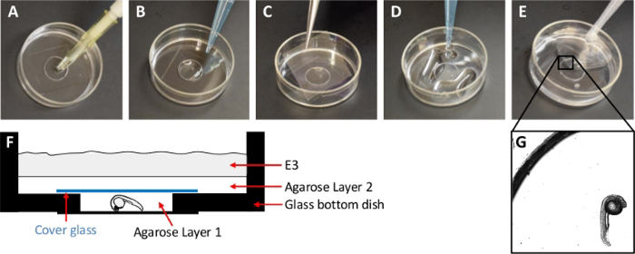
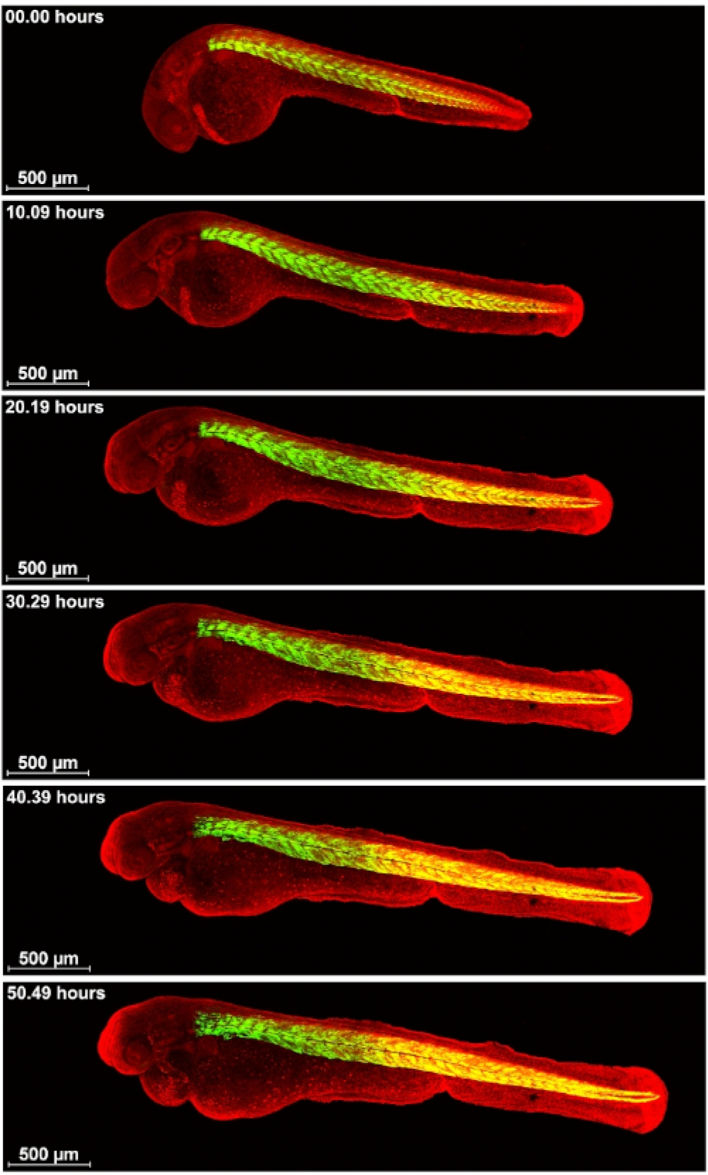
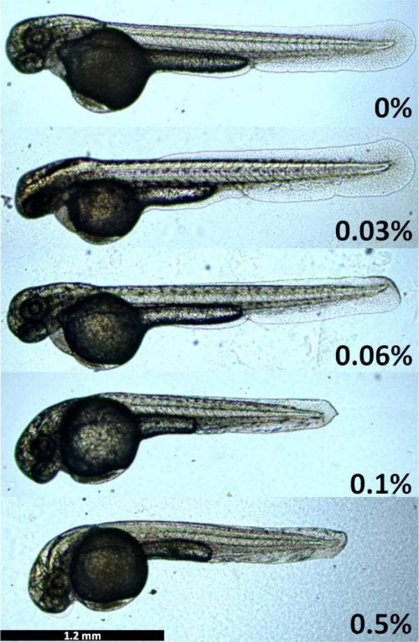
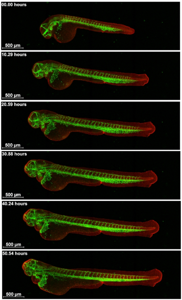
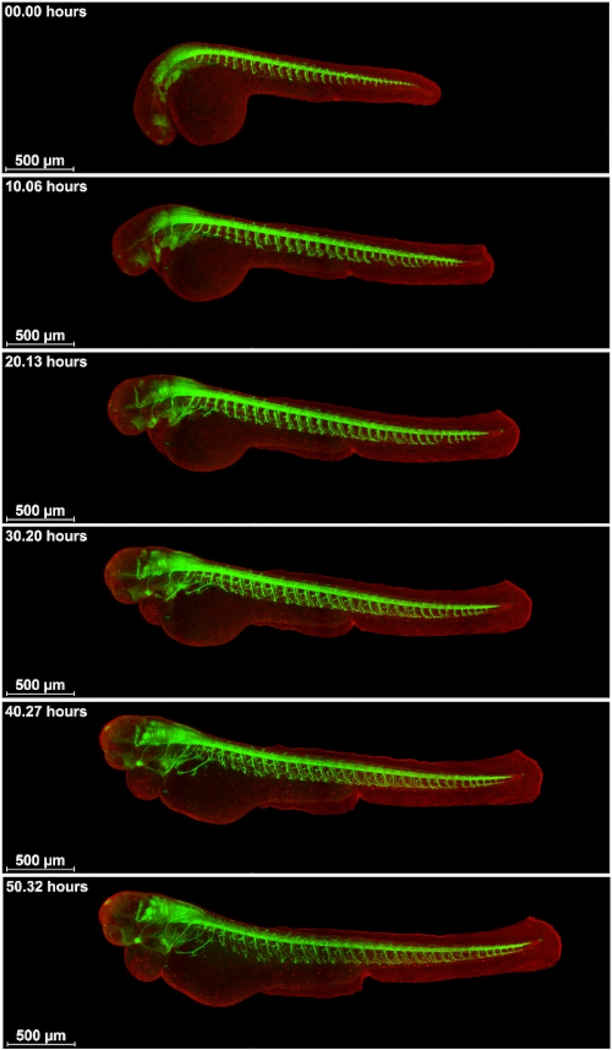
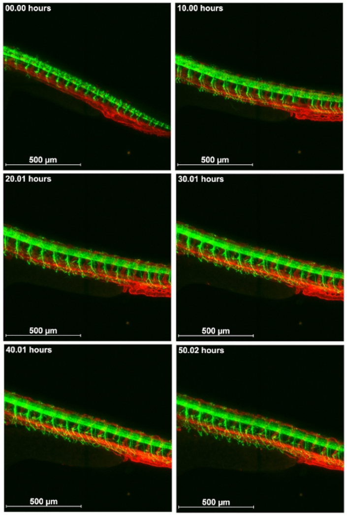
Dynamics of development can be followed by confocal time-lapse microscopy of live transgenic zebrafish embryos expressing fluorescence in specific tissues or cells. A difficulty with imaging whole embryo development is that zebrafish embryos grow substantially in length. When mounted as regularly done in 0.3-1% low melt agarose, the agarose imposes growth restriction, leading to distortions in the soft embryo body. Yet, to perform confocal time-lapse microscopy, the embryo must be immobilized. This article describes a layered mounting method for zebrafish embryos that restrict the motility of the embryos while allowing for the unrestricted growth. The mounting is performed in layers of agarose at different concentrations. To demonstrate the usability of this method, whole embryo vascular, neuronal and muscle development was imaged in transgenic fish for 55 consecutive hours. This mounting method can be used for easy, low-cost imaging of whole zebrafish embryos using inverted microscopes without requirements of molds or special equipment.

摘要

发育动力学可以通过对在特定组织或细胞中表达荧光的活转基因斑马鱼胚胎进行共聚焦延时显微镜观察来跟踪。对整个胚胎发育进行成像的一个困难在于斑马鱼胚胎的长度会大幅增长。当像常规那样将其固定在0.3 - 1%的低熔点琼脂糖中时,琼脂糖会限制生长,导致柔软的胚胎身体变形。然而,为了进行共聚焦延时显微镜观察,胚胎必须被固定。本文描述了一种用于斑马鱼胚胎的分层固定方法,该方法在限制胚胎运动的同时允许其不受限制地生长。固定是在不同浓度的琼脂糖层中进行的。为了证明该方法的可用性,对转基因鱼的整个胚胎血管、神经元和肌肉发育进行了连续55小时的成像。这种固定方法可用于使用倒置显微镜对整个斑马鱼胚胎进行简便、低成本的成像,无需模具或特殊设备。

https://cdn.ncbi.nlm.nih.gov/pmc/blobs/9421/8411746/107735cf9354/nihms-1735085-f0006.jpg
https://cdn.ncbi.nlm.nih.gov/pmc/blobs/9421/8411746/eeaf5569c0b9/nihms-1735085-f0001.jpg
https://cdn.ncbi.nlm.nih.gov/pmc/blobs/9421/8411746/d43d21e97620/nihms-1735085-f0002.jpg
https://cdn.ncbi.nlm.nih.gov/pmc/blobs/9421/8411746/572c848b1616/nihms-1735085-f0003.jpg
https://cdn.ncbi.nlm.nih.gov/pmc/blobs/9421/8411746/57f48908bc86/nihms-1735085-f0004.jpg
https://cdn.ncbi.nlm.nih.gov/pmc/blobs/9421/8411746/7a9403720d8c/nihms-1735085-f0005.jpg
https://cdn.ncbi.nlm.nih.gov/pmc/blobs/9421/8411746/107735cf9354/nihms-1735085-f0006.jpg

相似文献

1
A Layered Mounting Method for Extended Time-Lapse Confocal Microscopy of Whole Zebrafish Embryos.一种用于斑马鱼胚胎长时间延时共聚焦显微镜观察的分层固定方法。
J Vis Exp. 2020 Jan 14(155). doi: 10.3791/60321.
2
Analyzing craniofacial morphogenesis in zebrafish using 4D confocal microscopy.使用4D共聚焦显微镜分析斑马鱼的颅面形态发生。
J Vis Exp. 2014 Jan 30(83):e51190. doi: 10.3791/51190.
3
Fluorescence imaging of transgenic zebrafish embryos.转基因斑马鱼胚胎的荧光成像。
Cold Spring Harb Protoc. 2012 May 1;2012(5):pdb.prot069245. doi: 10.1101/pdb.prot069245.
4
Low-cost silicone imaging casts for zebrafish embryos and larvae.低成本硅胶成像铸型在斑马鱼胚胎和幼鱼中的应用。
Zebrafish. 2014 Feb;11(1):26-31. doi: 10.1089/zeb.2013.0897. Epub 2013 Nov 15.
5
Standardized mounting method of (zebrafish) embryos using a 3D-printed stamp for high-content, semi-automated confocal imaging.使用 3D 打印邮票对(斑马鱼)胚胎进行标准化装片,用于高通量、半自动化共聚焦成像。
BMC Biotechnol. 2019 Oct 22;19(1):68. doi: 10.1186/s12896-019-0558-y.
6
Time-lapse imaging beyond the diffraction limit.延时成像突破衍射极限。
Methods. 2018 Nov 1;150:32-41. doi: 10.1016/j.ymeth.2018.07.004. Epub 2018 Jul 27.
7
A Versatile Mounting Method for Long Term Imaging of Zebrafish Development.一种用于斑马鱼发育长期成像的通用固定方法。
J Vis Exp. 2017 Jan 26(119):55210. doi: 10.3791/55210.
8
Two-photon axotomy and time-lapse confocal imaging in live zebrafish embryos.斑马鱼活胚胎中的双光子轴突切断术和延时共聚焦成像。
J Vis Exp. 2009 Feb 16(24):1129. doi: 10.3791/1129.
9
Quantitative imaging of cell dynamics in mouse embryos using light-sheet microscopy.使用光片显微镜对小鼠胚胎中的细胞动力学进行定量成像。
Development. 2014 Nov;141(22):4406-14. doi: 10.1242/dev.111021. Epub 2014 Oct 24.
10
Analysis of Zebrafish Kidney Development with Time-lapse Imaging Using a Dissecting Microscope Equipped for Optical Sectioning.使用配备光学切片功能的解剖显微镜通过延时成像分析斑马鱼肾脏发育
J Vis Exp. 2016 Apr 7(110):e53921. doi: 10.3791/53921.

引用本文的文献

1
A method for creating custom 3D-printed molds to facilitate zebrafish imaging studies, including of cardiac development.一种创建定制3D打印模具以促进斑马鱼成像研究(包括心脏发育研究)的方法。
bioRxiv. 2025 Jun 3:2025.06.01.657246. doi: 10.1101/2025.06.01.657246.
2
A single-cell-resolution fate map of endoderm reveals demarcation of pancreatic progenitors by cell cycle.单细胞分辨率的内胚层命运图谱揭示了细胞周期对胰腺祖细胞的界定。
Proc Natl Acad Sci U S A. 2021 Jun 22;118(25). doi: 10.1073/pnas.2025793118.

本文引用的文献

1
A Versatile Mounting Method for Long Term Imaging of Zebrafish Development.一种用于斑马鱼发育长期成像的通用固定方法。
J Vis Exp. 2017 Jan 26(119):55210. doi: 10.3791/55210.
2
A Four-Well Dish for High-Resolution Longitudinal Imaging of the Tail and Posterior Trunk of Larval Zebrafish.一种用于高分辨率纵向成像斑马鱼幼体尾巴和后躯干的四孔培养皿。
Zebrafish. 2017 Oct;14(5):489-491. doi: 10.1089/zeb.2016.1406. Epub 2017 Jan 24.
3
Improved Long-Term Imaging of Embryos with Genetically Encoded α-Bungarotoxin.利用基因编码的α-银环蛇毒素改进胚胎的长期成像
PLoS One. 2015 Aug 5;10(8):e0134005. doi: 10.1371/journal.pone.0134005. eCollection 2015.
4
Generation of orientation tools for automated zebrafish screening assays using desktop 3D printing.使用桌面3D打印技术生成用于斑马鱼自动筛选试验的定向工具。
BMC Biotechnol. 2014 May 1;14:36. doi: 10.1186/1472-6750-14-36.
5
Embryonic exposure to sodium arsenite perturbs vascular development in zebrafish.胚胎期暴露于亚砷酸钠会扰乱斑马鱼的血管发育。
Aquat Toxicol. 2014 Jul;152:152-63. doi: 10.1016/j.aquatox.2014.04.006. Epub 2014 Apr 16.
6
Low-cost silicone imaging casts for zebrafish embryos and larvae.低成本硅胶成像铸型在斑马鱼胚胎和幼鱼中的应用。
Zebrafish. 2014 Feb;11(1):26-31. doi: 10.1089/zeb.2013.0897. Epub 2013 Nov 15.
7
High-speed panoramic light-sheet microscopy reveals global endodermal cell dynamics.高速全景光片显微镜揭示了全局内胚层细胞动力学。
Nat Commun. 2013;4:2207. doi: 10.1038/ncomms3207.
8
Zebrabow: multispectral cell labeling for cell tracing and lineage analysis in zebrafish.斑马鱼:多光谱细胞标记用于细胞示踪和谱系分析。
Development. 2013 Jul;140(13):2835-46. doi: 10.1242/dev.094631.
9
Regular care and maintenance of a zebrafish (Danio rerio) laboratory: an introduction.斑马鱼(Danio rerio)实验室的定期护理与维护:引言
J Vis Exp. 2012 Nov 18(69):e4196. doi: 10.3791/4196.
10
Multilayer mounting enables long-term imaging of zebrafish development in a light sheet microscope.多层挂载可实现斑马鱼发育的光片显微镜的长期成像。
Development. 2012 Sep;139(17):3242-7. doi: 10.1242/dev.082586.