Jafri Mohammad Alam, Kalamegam Gauthaman, Abbas Mohammed, Al-Kaff Mohammed, Ahmed Farid, Bakhashab Sherin, Rasool Mahmood, Naseer Muhammad Imran, Sinnadurai Vasan, Pushparaj Peter Natesan
Centre of Excellence in Genomic Medicine Research, King Abdulaziz University, Jeddah, Saudi Arabia.
Department of Medical Laboratory Technology, Faculty of Applied Medical Sciences, King Abdulaziz University, Jeddah, Saudi Arabia.
Front Cell Dev Biol. 2020 Jan 17;7:380. doi: 10.3389/fcell.2019.00380. eCollection 2019.
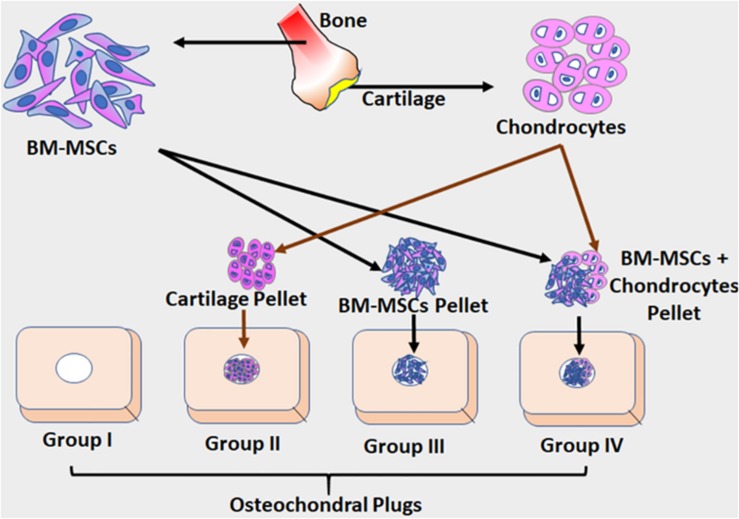
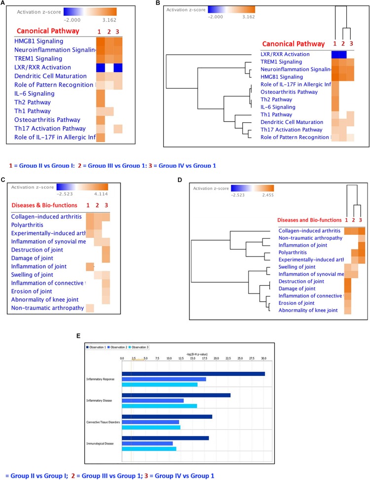
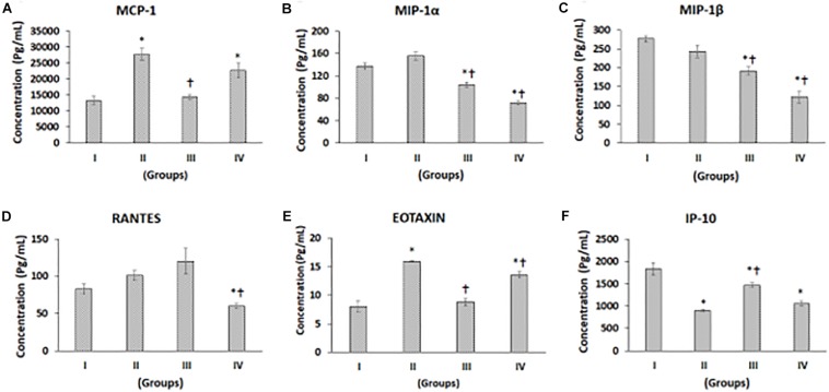
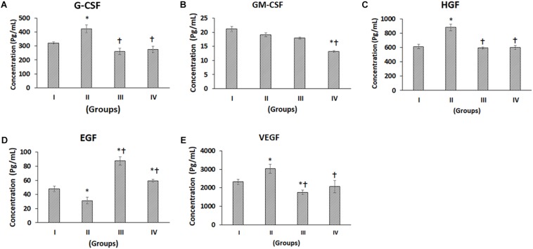
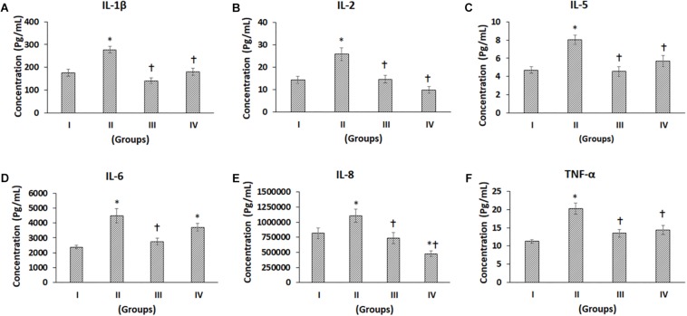
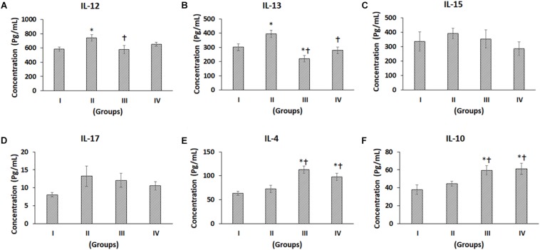
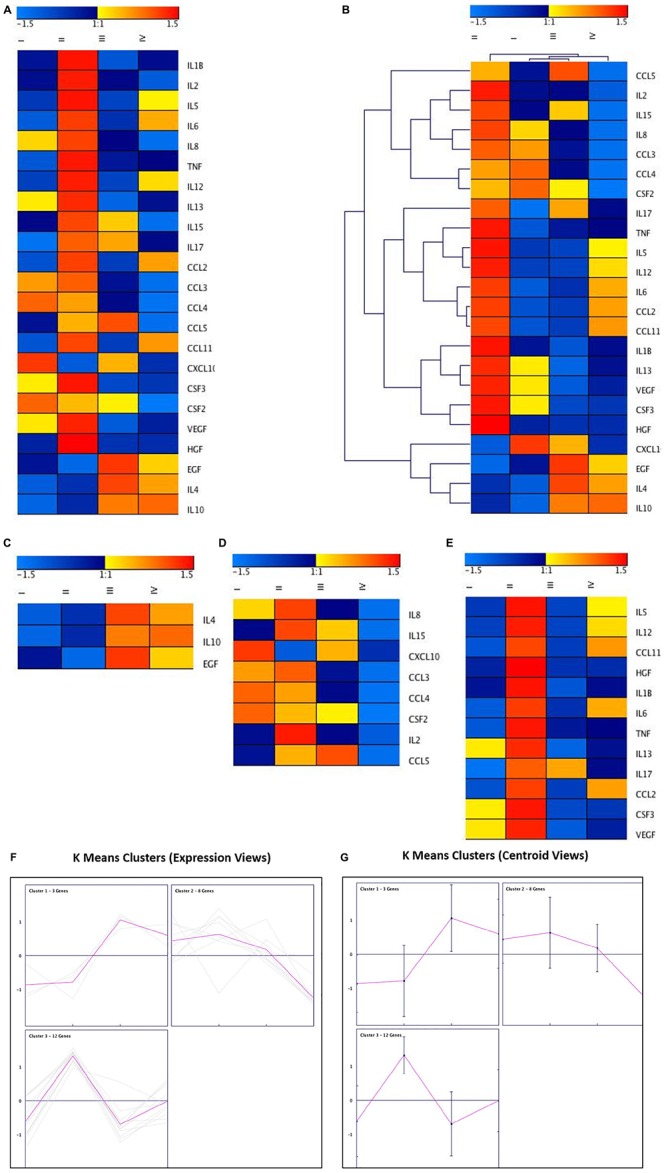
Osteoarthritis (OA) is a chronic degenerative joint disorder associated with degradation and decreased production of the extracellular matrix, eventually leading to cartilage destruction. Limited chondrocyte turnover, structural damage, and prevailing inflammatory milieu prevent efficient cartilage repair and restoration of joint function. In the present study, we evaluated the role of secreted cytokines, chemokines, and growth factors present in the culture supernatant obtained from an osteochondral model of cartilage differentiation using cartilage pellets (CP), bone marrow stem cells (BM-MSCs), and/or BM-MSCs + CP. Multiplex cytokine analysis showed differential secretion of growth factors (G-CSF, GM-CSF, HGF, EGF, VEGF); chemokines (MCP-1, MIP1α, MIP1β, RANTES, Eotaxin, IP-10), pro-inflammatory cytokines (IL-1β, IL-2, IL-5, IL-6, IL-8, TNFα, IL-12, IL-15, IL-17) and anti-inflammatory cytokines (IL-4, IL-10, and IL-13) in the experimental groups compared to the control. analyses of the role of stem cells and CP in relation to the expression of various molecules, canonical pathways and hierarchical cluster patterns were deduced using the Ingenuity Pathway Analysis (IPA) software (Qiagen, United States). The interactions of the cytokines, chemokines, and growth factors that are involved in the cartilage differentiation showed that stem cells, when used together with CP, bring about a favorable cell signaling that supports cartilage differentiation and additionally helps to attenuate inflammatory cytokines and further downstream disease-associated pro-inflammatory pathways. Hence, the autologous or allogeneic stem cells and local cartilage tissues may be used for efficient cartilage differentiation and the management of OA.
骨关节炎(OA)是一种慢性退行性关节疾病,与细胞外基质的降解和生成减少相关,最终导致软骨破坏。有限的软骨细胞更新、结构损伤和普遍存在的炎症环境阻碍了软骨的有效修复和关节功能的恢复。在本研究中,我们评估了使用软骨微球(CP)、骨髓干细胞(BM-MSCs)和/或BM-MSCs + CP从软骨分化的骨软骨模型获得的培养上清液中分泌的细胞因子、趋化因子和生长因子的作用。多重细胞因子分析显示,与对照组相比,实验组中生长因子(G-CSF、GM-CSF、HGF、EGF、VEGF)、趋化因子(MCP-1、MIP1α、MIP1β、RANTES、嗜酸性粒细胞趋化因子、IP-10)、促炎细胞因子(IL-1β、IL-2、IL-5、IL-6、IL-8、TNFα、IL-12、IL-15、IL-17)和抗炎细胞因子(IL-4、IL-10和IL-13)的分泌存在差异。使用 Ingenuity Pathway Analysis(IPA)软件(美国Qiagen公司)推导了干细胞和CP在各种分子表达、经典途径和层次聚类模式方面的作用分析。参与软骨分化的细胞因子、趋化因子和生长因子的相互作用表明,干细胞与CP一起使用时,会产生有利的细胞信号传导,支持软骨分化,并有助于减弱炎症细胞因子和进一步下游与疾病相关的促炎途径。因此,自体或异体干细胞以及局部软骨组织可用于有效的软骨分化和骨关节炎的管理。