Department of Physiological Science and Molecular Biology, Fukuoka Dental College, Fukuoka, Japan.
Department of Oral Maxillofacial Surgery, Fukuoka Dental College, Fukuoka, Japan.
Cancer Sci. 2020 Apr;111(4):1113-1123. doi: 10.1111/cas.14336. Epub 2020 Feb 29.
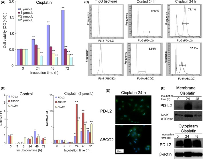
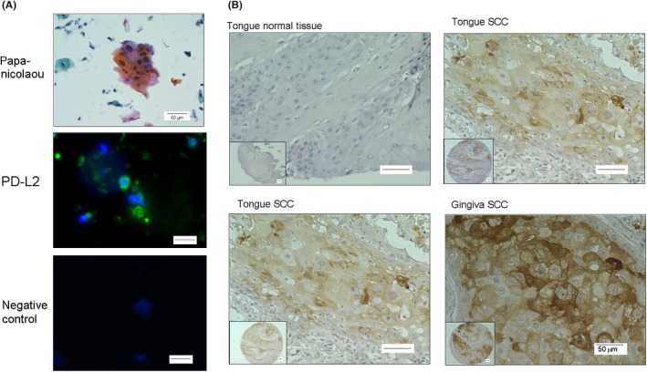
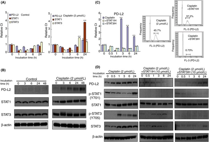
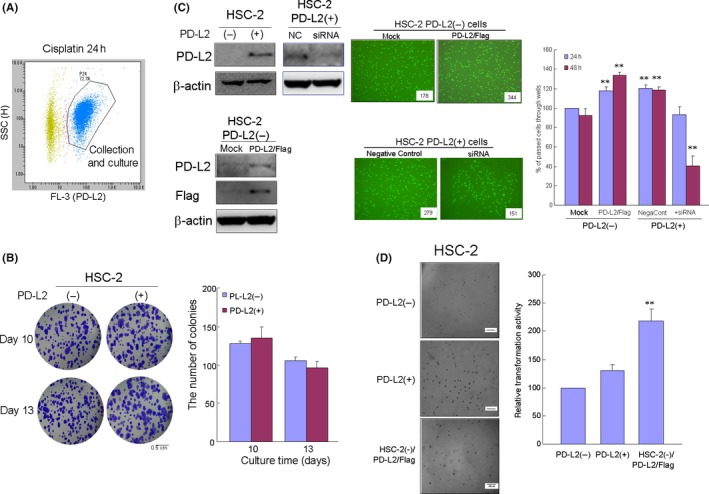
Programmed cell death ligands (PD-Ls) are expressed in tumor cells where they bind to programmed cell death-1, an immunocyte co-receptor, resulting in tumor cell evasion from the immune system. Chemotherapeutic drugs have been recently reported to induce the expression of PD-L, such as PD-L1, in some cancer cells. However, little is known regarding PD-L2 expression and its role in oral squamous cell carcinoma (OSCC). In this study, we examined the effect of cisplatin on the expression and regulation of PD-L2 in OSCC cell lines and analyzed malignant behavior in PD-L2-expressing cells using colony, transwell and transformation assays. In addition, we examined PD-L2 expression in the tumor tissues of OSCC patients using cytology and tissue microarray methods. In OSCC cell lines, cisplatin treatment upregulated PD-L2 expression, along with that of the drug efflux transporter ABCG2, via signal transducers and activator of transcription (STAT) 1/3 activation. Moreover, PD-L2-positive or PD-L2-overexpressing cells demonstrated upregulation in both invasion and transformation ability but not in proliferation compared with PD-L2-negative or PD-L2-silencing cells. PD-L2 expression was also observed in OSCC cells of cytology samples and tissue from OSCC patients. The intensity of PD-L2 expression was correlated with more malignant morphological features in the histological appearance and an invasive pattern. Our findings indicate that cisplatin-upregulated PD-L2 expression in OSCC via STAT1/3 activation and the expression of PD-L2 are likely to be associated with malignancy in OSCC. The PD-L2 expression in cisplatin-resistant OSCC cells may be a critical factor in prognosis of advanced OSCC patients.
程序性细胞死亡配体(PD-Ls)在肿瘤细胞中表达,它们与程序性细胞死亡受体 1(一种免疫细胞共受体)结合,导致肿瘤细胞逃避免疫系统。最近有报道称,化疗药物可诱导某些癌细胞中 PD-L 的表达,如 PD-L1。然而,关于 PD-L2 的表达及其在口腔鳞状细胞癌(OSCC)中的作用知之甚少。在这项研究中,我们研究了顺铂对 OSCC 细胞系中 PD-L2 表达的影响及其调控机制,并通过集落、Transwell 和转化实验分析了 PD-L2 表达细胞的恶性行为。此外,我们还通过细胞学和组织微阵列方法检查了 OSCC 患者肿瘤组织中的 PD-L2 表达。在 OSCC 细胞系中,顺铂处理通过信号转导和转录激活因子(STAT)1/3 激活上调 PD-L2 表达及其药物外排转运蛋白 ABCG2 的表达。此外,与 PD-L2 阴性或 PD-L2 沉默细胞相比,PD-L2 阳性或 PD-L2 过表达细胞的侵袭和转化能力上调,但增殖能力没有上调。在细胞学样本的 OSCC 细胞和 OSCC 患者的组织中也观察到 PD-L2 的表达。PD-L2 表达的强度与组织学外观和侵袭模式中更恶性的形态特征相关。我们的研究结果表明,顺铂通过 STAT1/3 激活上调 OSCC 中的 PD-L2 表达,PD-L2 的表达可能与 OSCC 的恶性程度有关。顺铂耐药 OSCC 细胞中 PD-L2 的表达可能是晚期 OSCC 患者预后的一个关键因素。