Service de Pneumologie et d'Allergologie Pédiatriques, AP-HP, Hôpital Universitaire Necker-Enfants Malades, 75743 Cedex 15, Paris, France.
INSERM, U955, Institut Mondor de Recherche Biomedicale (IMRB), Equipe 4, 94000, Créteil, France.
Respir Res. 2020 Feb 4;21(1):43. doi: 10.1186/s12931-020-1306-5.
The pathophysiology of congenital cystic adenomatoid malformations (CCAM) of the lung remains poorly understood.
This study aimed to identify more precisely the molecular mechanisms limited to a compartment of lung tissue, through a transcriptomic analysis of the epithelium of macrocystic forms.
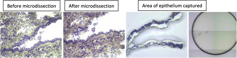
Tissue fragments displaying CCAM were obtained during planned surgical resections. Epithelial mRNA was obtained from cystic and normal areas after laser capture microdissection (LCM). Transcriptomic analyses were performed and the results were confirmed by RT-PCR and immunohistochemistry in independent samples.
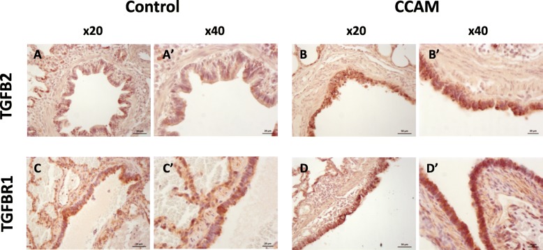
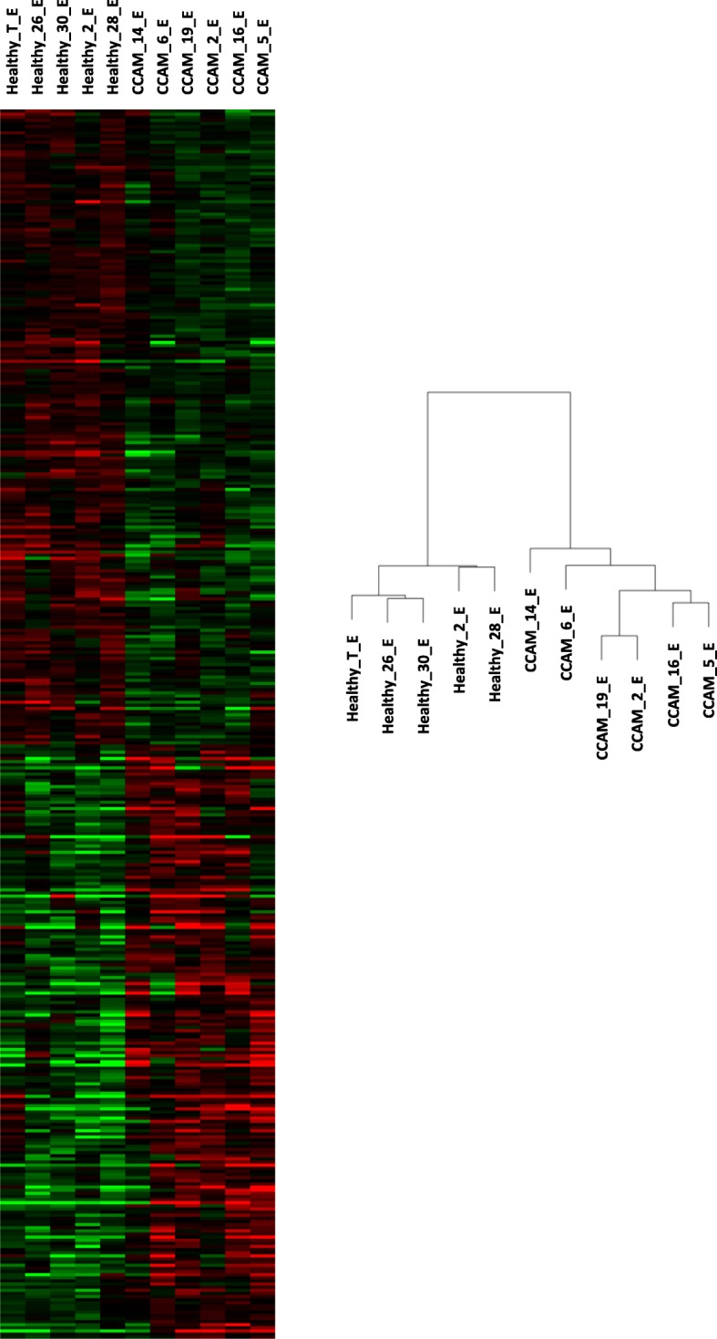
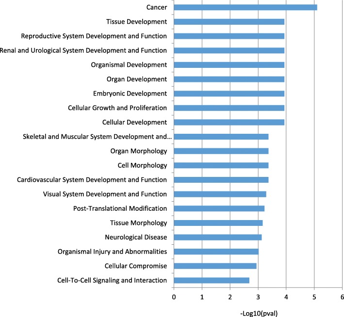
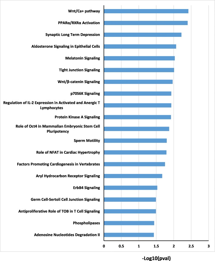
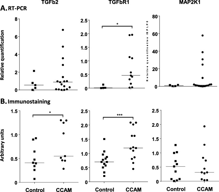
After controlling for RNA quality, we analysed the transcriptomes of six cystic areas and five control areas. In total, 393 transcripts were differentially expressed in the epithelium, between CCAM and control areas. The most highly redundant genes involved in biological functions and signalling pathways differentially expressed between CCAM and control epithelium included TGFB2, TGFBR1, and MAP 2 K1. These genes were considered particularly relevant as they have been implicated in branching morphogenesis. RT-qPCR analysis confirmed in independent samples that TGFBR1 was more strongly expressed in CCAM than in control tissues (p < 0.03). Immunohistochemistry analysis showed TGFBR1 (p = 0.0007) and TGFB2 (p < 0.02) levels to be significantly higher in the epithelium of CCAM than in that of control tissues.
This compartmentalised transcriptomic analysis of the epithelium of macrocystic lung malformations identified a dysregulation of TGFB signalling at the mRNA and protein levels, suggesting a possible role of this pathway in CCAM pathogenesis.
ClinicalTrials.gov Identifier: NCT01732185.
先天性囊性腺瘤样畸形(CCAM)的病理生理学仍知之甚少。
本研究旨在通过对大囊型肺组织的上皮细胞进行转录组分析,更精确地确定仅局限于肺组织某一部位的分子机制。
在计划的手术切除过程中获得显示 CCAM 的组织片段。使用激光捕获显微切割(LCM)从囊状和正常区域获得上皮 mRNA。进行转录组分析,并在独立样本中通过 RT-PCR 和免疫组织化学验证结果。
在控制 RNA 质量后,我们分析了六个囊状区域和五个对照区域的转录组。总共,CCAM 与对照上皮组织之间的上皮细胞中差异表达了 393 个转录本。差异表达最多的与生物功能和信号通路相关的基因包括 TGFB2、TGFBR1 和 MAP2K1。这些基因被认为特别重要,因为它们与分支形态发生有关。在独立样本中,RT-qPCR 分析证实 TGFBR1 在 CCAM 中的表达强于对照组织(p<0.03)。免疫组织化学分析显示,TGFBR1(p=0.0007)和 TGFB2(p<0.02)在 CCAM 上皮组织中的水平明显高于对照组织。
对大囊型肺畸形的上皮细胞进行的这种分区转录组分析发现,TGFB 信号在 mRNA 和蛋白水平上失调,提示该途径可能在 CCAM 发病机制中起作用。
ClinicalTrials.gov 标识符:NCT01732185。