• 文献检索
  • 文档翻译
  • 深度研究
  • 学术资讯
  • Suppr Zotero 插件Zotero 插件
  • 邀请有礼
  • 套餐&价格
  • 历史记录
应用&插件
Suppr Zotero 插件Zotero 插件浏览器插件Mac 客户端Windows 客户端微信小程序
定价
高级版会员购买积分包购买API积分包
服务
文献检索文档翻译深度研究API 文档MCP 服务
关于我们
关于 Suppr公司介绍联系我们用户协议隐私条款
关注我们

Suppr 超能文献

核心技术专利:CN118964589B侵权必究
粤ICP备2023148730 号-1Suppr @ 2026

文献检索

告别复杂PubMed语法,用中文像聊天一样搜索,搜遍4000万医学文献。AI智能推荐,让科研检索更轻松。

立即免费搜索

文件翻译

保留排版,准确专业,支持PDF/Word/PPT等文件格式,支持 12+语言互译。

免费翻译文档

深度研究

AI帮你快速写综述,25分钟生成高质量综述,智能提取关键信息,辅助科研写作。

立即免费体验

纳米容器处理的巨噬细胞的多模式 X 射线成像和骨陷窝周围骨基质中的钙分布。

Multimodal X-ray imaging of nanocontainer-treated macrophages and calcium distribution in the perilacunar bone matrix.

机构信息

DESY Photon Science, Deutsches Elektronen-Synchrotron DESY, Hamburg, 22607, Germany.

Center for Free-Electron Laser Science, Hamburg, 22607, Germany.

出版信息

Sci Rep. 2020 Feb 4;10(1):1784. doi: 10.1038/s41598-020-58318-7.

DOI:10.1038/s41598-020-58318-7
PMID:32019946
原文链接:https://pmc.ncbi.nlm.nih.gov/articles/PMC7000813/
Abstract

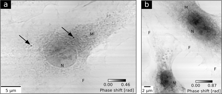
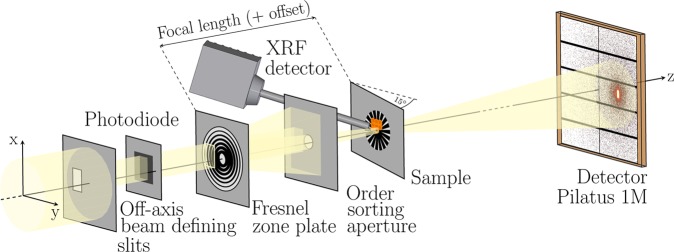
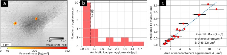
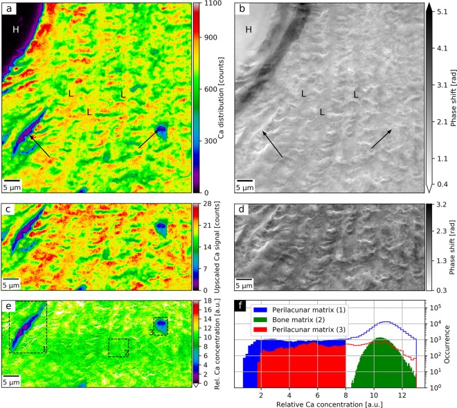
Studies of biological systems typically require the application of several complementary methods able to yield statistically-relevant results at a unique level of sensitivity. Combined X-ray fluorescence and ptychography offer excellent elemental and structural imaging contrasts at the nanoscale. They enable a robust correlation of elemental distributions with respect to the cellular morphology. Here we extend the applicability of the two modalities to higher X-ray excitation energies, permitting iron mapping. Using a long-range scanning setup, we applied the method to two vital biomedical cases. We quantified the iron distributions in a population of macrophages treated with Mycobacterium-tuberculosis-targeting iron-oxide nanocontainers. Our work allowed to visualize the internalization of the nanocontainer agglomerates in the cytosol. From the iron areal mass maps, we obtained a distribution of antibiotic load per agglomerate and an average areal concentration of nanocontainers in the agglomerates. In the second application we mapped the calcium content in a human bone matrix in close proximity to osteocyte lacunae (perilacunar matrix). A concurrently acquired ptychographic image was used to remove the mass-thickness effect from the raw calcium map. The resulting ptychography-enhanced calcium distribution allowed then to observe a locally lower degree of mineralization of the perilacunar matrix.

摘要

研究生物系统通常需要应用几种互补的方法,这些方法能够在独特的灵敏度水平上产生具有统计学意义的结果。X 射线荧光和相衬技术的联合应用在纳米尺度上提供了极好的元素和结构成像对比。它们能够实现元素分布与细胞形态之间的稳健相关性。在这里,我们将这两种模式的适用性扩展到更高的 X 射线激发能量,从而允许进行铁成像。使用远程扫描设置,我们将该方法应用于两个重要的生物医学案例。我们定量分析了用针对结核分枝杆菌的氧化铁纳米容器处理的巨噬细胞群体中的铁分布。我们的工作使纳米容器聚集体在细胞质中的内化可视化。从铁的面质量图中,我们获得了每个聚集体中的抗生素负载分布和聚集体中纳米容器的平均面浓度。在第二个应用中,我们绘制了与人骨基质中骨陷窝(骨周基质)附近的钙含量的分布图。同时获得的相衬图像用于从原始钙图中去除质量厚度效应。由此产生的相衬增强钙分布使得能够观察到骨周基质局部矿化程度较低。

https://cdn.ncbi.nlm.nih.gov/pmc/blobs/858a/7000813/723ae2fc1cd6/41598_2020_58318_Fig4_HTML.jpg
https://cdn.ncbi.nlm.nih.gov/pmc/blobs/858a/7000813/60f0bfb917da/41598_2020_58318_Fig1_HTML.jpg
https://cdn.ncbi.nlm.nih.gov/pmc/blobs/858a/7000813/1ca7e6adabb6/41598_2020_58318_Fig2_HTML.jpg
https://cdn.ncbi.nlm.nih.gov/pmc/blobs/858a/7000813/45ac1ef429fa/41598_2020_58318_Fig3_HTML.jpg
https://cdn.ncbi.nlm.nih.gov/pmc/blobs/858a/7000813/723ae2fc1cd6/41598_2020_58318_Fig4_HTML.jpg

相似文献

1
Multimodal X-ray imaging of nanocontainer-treated macrophages and calcium distribution in the perilacunar bone matrix.纳米容器处理的巨噬细胞的多模式 X 射线成像和骨陷窝周围骨基质中的钙分布。
Sci Rep. 2020 Feb 4;10(1):1784. doi: 10.1038/s41598-020-58318-7.
2
Glucocorticoid suppression of osteocyte perilacunar remodeling is associated with subchondral bone degeneration in osteonecrosis.糖皮质激素抑制破骨细胞陷窝周重塑与骨坏死的软骨下骨退变有关。
Sci Rep. 2017 Mar 22;7:44618. doi: 10.1038/srep44618.
3
3D osteocyte lacunar morphometric properties and distributions in human femoral cortical bone using synchrotron radiation micro-CT images.利用同步辐射微计算机断层扫描图像研究人股骨皮质骨中 3D 骨细胞陷窝的形态计量学特征及其分布
Bone. 2014 Mar;60:172-85. doi: 10.1016/j.bone.2013.12.008. Epub 2013 Dec 12.
4
Vitamin D regulates osteocyte survival and perilacunar remodeling in human and murine bone.维生素 D 调节人骨和鼠骨中破骨细胞的存活和骨陷窝周围重塑。
Bone. 2017 Oct;103:78-87. doi: 10.1016/j.bone.2017.06.022. Epub 2017 Jun 27.
5
Bone Matrix Maturation in a Rat Model of Intra-Cortical Bone Remodeling.皮质内骨重塑大鼠模型中的骨基质成熟
Calcif Tissue Int. 2017 Aug;101(2):193-203. doi: 10.1007/s00223-017-0270-7. Epub 2017 Apr 3.
6
Osteocytic perilacunar/canalicular turnover in hemodialysis patients with high and low serum PTH levels.高和低血清甲状旁腺激素水平血液透析患者的骨细胞性骨陷窝/骨小管更新。
Bone. 2018 Aug;113:68-76. doi: 10.1016/j.bone.2018.05.002. Epub 2018 May 5.
7
Physiological and pathological osteocytic osteolysis.生理性和病理性骨细胞性骨溶解
J Musculoskelet Neuronal Interact. 2018 Sep 1;18(3):292-303.
8
PTH signaling mediates perilacunar remodeling during exercise.甲状旁腺激素信号传导在运动过程中介导腔周重塑。
Matrix Biol. 2016 May-Jul;52-54:162-175. doi: 10.1016/j.matbio.2016.02.010. Epub 2016 Feb 23.
9
Parallel mechanisms suppress cochlear bone remodeling to protect hearing.并联机制抑制耳蜗骨重塑以保护听力。
Bone. 2016 Aug;89:7-15. doi: 10.1016/j.bone.2016.04.010. Epub 2016 Apr 13.
10
Increased proportion of hypermineralized osteocyte lacunae in osteoporotic and osteoarthritic human trabecular bone: implications for bone remodeling.骨质疏松症和骨关节炎患者松质骨中海锦骨陷窝矿化过度比例增加:对骨重建的影响。
Bone. 2012 Mar;50(3):688-94. doi: 10.1016/j.bone.2011.11.021. Epub 2011 Dec 7.

引用本文的文献

1
Dose-efficient multimodal microscopy of human tissue at a hard X-ray nanoprobe beamline.在硬 X 射线纳米探针光束线上实现高效的人组织多模式显微镜技术。
J Synchrotron Radiat. 2022 May 1;29(Pt 3):807-815. doi: 10.1107/S1600577522001874. Epub 2022 Mar 16.
2
First ptychographic X-ray computed tomography experiment on the NanoMAX beamline.在NanoMAX光束线上进行的首次叠层X射线计算机断层扫描实验。
J Appl Crystallogr. 2020 Oct 13;53(Pt 6):1444-1451. doi: 10.1107/S160057672001211X. eCollection 2020 Dec 1.
3
PtyNAMi: ptychographic nano-analytical microscope.

本文引用的文献

1
Correlative 3D x-ray fluorescence and ptychographic tomography of frozen-hydrated green algae.冷冻水合绿藻的相关 3D X 射线荧光和叠层相位衬度断层成像。
Sci Adv. 2018 Nov 2;4(11):eaau4548. doi: 10.1126/sciadv.aau4548. eCollection 2018 Nov.
2
Design and performance of an X-ray scanning microscope at the Hard X-ray Nanoprobe beamline of NSLS-II.NSLS-II硬X射线纳米探针光束线处的X射线扫描显微镜的设计与性能
J Synchrotron Radiat. 2017 Nov 1;24(Pt 6):1113-1119. doi: 10.1107/S1600577517011183. Epub 2017 Oct 5.
3
Osteocytes Acidify Their Microenvironment in Response to PTHrP In Vitro and in Lactating Mice In Vivo.
PtyNAMi:叠层成像纳米分析显微镜。
J Appl Crystallogr. 2020 Jul 30;53(Pt 4):957-971. doi: 10.1107/S1600576720008420. eCollection 2020 Aug 1.
骨细胞在体外对甲状旁腺激素相关蛋白(PTHrP)以及在哺乳期小鼠体内做出反应时会酸化其微环境。
J Bone Miner Res. 2017 Aug;32(8):1761-1772. doi: 10.1002/jbmr.3167. Epub 2017 Jun 12.
4
Combined in-situ imaging of structural organization and elemental composition of substantia nigra neurons in the elderly.老年人黑质神经元结构组织与元素组成的联合原位成像
Talanta. 2016 Dec 1;161:368-376. doi: 10.1016/j.talanta.2016.08.023. Epub 2016 Aug 5.
5
Multifunctional phosphate-based inorganic-organic hybrid nanoparticles.多功能磷酸盐基无机-有机杂化纳米粒子。
J Am Chem Soc. 2015 Jun 17;137(23):7329-36. doi: 10.1021/jacs.5b01172. Epub 2015 Jun 8.
6
Element-specific X-ray phase tomography of 3D structures at the nanoscale.纳米尺度 3D 结构的元素特定 X 射线相层析成像。
Phys Rev Lett. 2015 Mar 20;114(11):115501. doi: 10.1103/PhysRevLett.114.115501. Epub 2015 Mar 16.
7
Macrophage defense mechanisms against intracellular bacteria.巨噬细胞针对细胞内细菌的防御机制。
Immunol Rev. 2015 Mar;264(1):182-203. doi: 10.1111/imr.12266.
8
Simultaneous cryo X-ray ptychographic and fluorescence microscopy of green algae.绿藻的同步冷冻X射线叠层成像和荧光显微镜观察
Proc Natl Acad Sci U S A. 2015 Feb 24;112(8):2314-9. doi: 10.1073/pnas.1413003112. Epub 2015 Feb 9.
9
Intravital bone imaging by two-photon excitation microscopy to identify osteocytic osteolysis in vivo.通过双光子激发显微镜进行活体骨成像以在体内识别骨细胞性骨溶解。
Bone. 2015 May;74:134-9. doi: 10.1016/j.bone.2015.01.013. Epub 2015 Jan 24.
10
Continuous scanning mode for ptychography.叠层成像术的连续扫描模式。
Opt Lett. 2014 Oct 15;39(20):6066-9. doi: 10.1364/OL.39.006066.