Stem Cell Program, Boston Children's Hospital, Boston, MA, USA.
Department of Stem Cell and Regenerative Biology, Harvard University, Cambridge, MA, USA.
Nature. 2020 Feb;578(7794):278-283. doi: 10.1038/s41586-020-1971-z. Epub 2020 Feb 5.
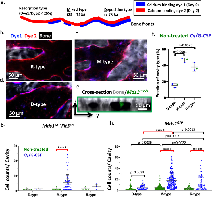
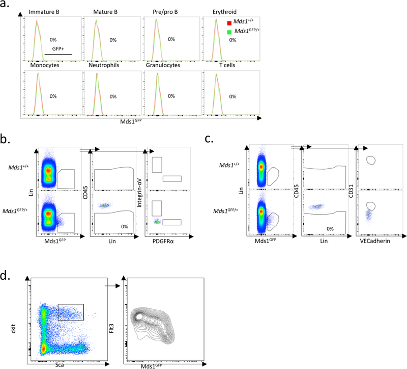
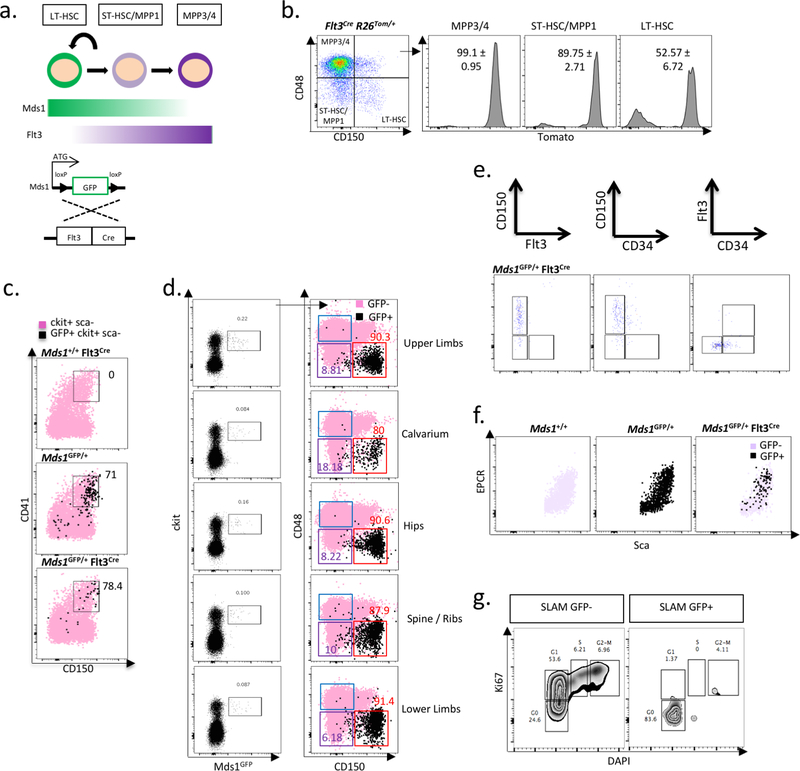
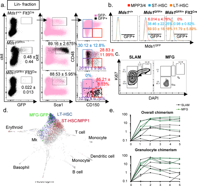
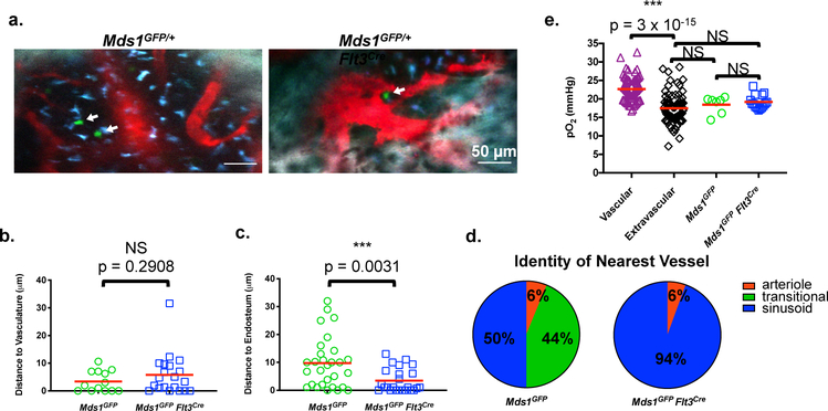
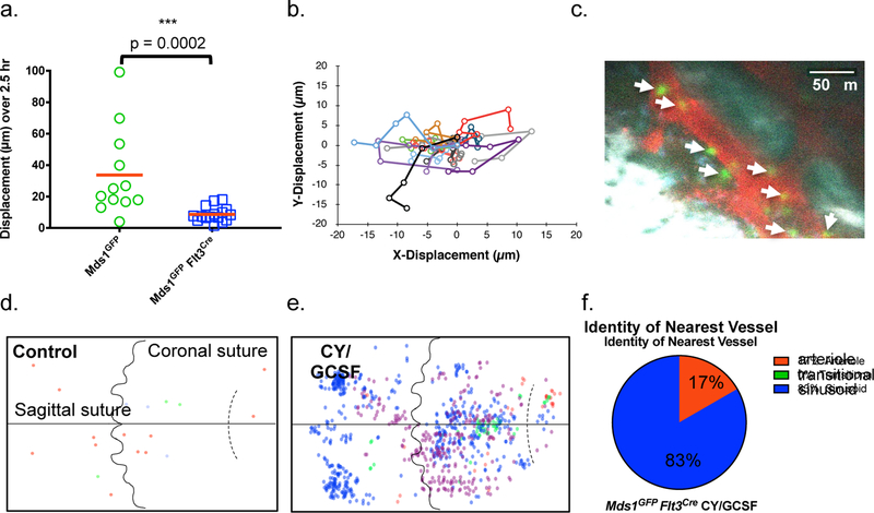
The biology of haematopoietic stem cells (HSCs) has predominantly been studied under transplantation conditions. It has been particularly challenging to study dynamic HSC behaviour, given that the visualization of HSCs in the native niche in live animals has not, to our knowledge, been achieved. Here we describe a dual genetic strategy in mice that restricts reporter labelling to a subset of the most quiescent long-term HSCs (LT-HSCs) and that is compatible with current intravital imaging approaches in the calvarial bone marrow. We show that this subset of LT-HSCs resides close to both sinusoidal blood vessels and the endosteal surface. By contrast, multipotent progenitor cells (MPPs) show greater variation in distance from the endosteum and are more likely to be associated with transition zone vessels. LT-HSCs are not found in bone marrow niches with the deepest hypoxia and instead are found in hypoxic environments similar to those of MPPs. In vivo time-lapse imaging revealed that LT-HSCs at steady-state show limited motility. Activated LT-HSCs show heterogeneous responses, with some cells becoming highly motile and a fraction of HSCs expanding clonally within spatially restricted domains. These domains have defined characteristics, as HSC expansion is found almost exclusively in a subset of bone marrow cavities with bone-remodelling activity. By contrast, cavities with low bone-resorbing activity do not harbour expanding HSCs. These findings point to previously unknown heterogeneity within the bone marrow microenvironment, imposed by the stages of bone turnover. Our approach enables the direct visualization of HSC behaviours and dissection of heterogeneity in HSC niches.
造血干细胞(HSCs)的生物学主要在移植条件下进行研究。由于尚未实现 HSCs 在天然龛位中的动态行为的可视化,因此研究动态 HSC 行为尤其具有挑战性。在这里,我们在小鼠中描述了一种双重遗传策略,该策略将报告基因标记限制在最静止的长期 HSCs(LT-HSCs)亚群中,并且与颅骨髓内目前的活体成像方法兼容。我们表明,LT-HSCs 的这一部分位于窦状血管和骨内膜表面附近。相比之下,多能祖细胞(MPPs)与骨内膜的距离变化更大,并且更有可能与过渡区血管相关。LT-HSCs 不在骨髓龛中具有最深的缺氧,而是在类似于 MPPs 的缺氧环境中。体内延时成像显示,稳态下的 LT-HSCs 运动性有限。激活的 LT-HSCs 表现出异质性反应,一些细胞变得高度运动,一部分 HSCs 在空间受限的区域内克隆性扩增。这些区域具有确定的特征,因为 HSC 扩增几乎仅发生在具有骨重塑活性的一部分骨髓腔中。相比之下,具有低骨吸收活性的腔不含有扩增的 HSCs。这些发现表明,骨转换阶段对骨髓微环境内的 HSCs 龛位施加了以前未知的异质性。我们的方法能够直接观察 HSC 行为并剖析 HSC 龛位的异质性。