From the Pathology and Laboratory Medicine Department, College of Medicine, King Abdulaziz Medical City, Riyadh, Saudi Arabia.
From the College of Medicine, King Saud bin Abdulaziz University for Health Sciences, Riyadh, Saudi Arabia.
Ann Saudi Med. 2020 Jan-Feb;40(1):36-41. doi: 10.5144/0256-4947.2020.36. Epub 2020 Feb 6.
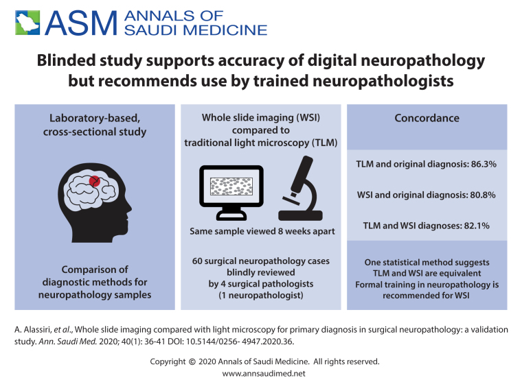
Digital pathology practice is rapidly gaining popularity among practicing anatomic pathologists. Acceptance is higher among the newer generation of pathologists who are willing to adapt to this new diagnostic method due to the advantages offered by whole slide imaging (WSI) compared to traditional light microscopy (TLM). We performed this validation study because we plan to implement the WSI system for diagnostic services.
Determine the feasibility of using digital pathology for diagnostic services by assessing the equivalency of WSI and TLM.
A laboratory-based cross-sectional study.
Central laboratory at a tertiary health care center.
Four practicing surgical pathologists participated in this study. Each pathologist blindly reviewed 60 surgical neuropathology cases with a minimum 8-week washout-period between the two diagnostic modalities (WSI vs. TLM). Intraobserver concordance rates between WSI and TLM diagnoses as compared to the original diagnosis were calculated.
Overall intraobserver concordance rates between each diagnostic method (WSI and TLM) and original diagnosis.
60 in-house surgical neuropathology cases.
The overall intraobserver concordance rate between TLM and original diagnosis was 86.3% (range 76.7%-91.7%) versus 80.8% for WSI (range 68.3%-88.3%). These findings are suggestive of the superiority of TLM, but the Fleiss' Kappa statistic indicated that the two methods are equivalent, despite the low level of the K value.
WSI is not inferior to the light microscopy and is feasible for primary diagnosis in surgical neuropathology. However, to ensure the best results, only formally trained neuropathologists should handle the digital neuropathology service.
Only one diagnostic slide per case rather than the whole set of slides, sample size was relatively small, and there was an insufficient number of participating neuropathologists.
None.
数字病理学实践在解剖病理学家中迅速流行。由于全玻片成像(WSI)相对于传统光学显微镜(TLM)具有优势,新一代愿意适应这种新诊断方法的病理学家对其接受程度更高。我们进行了这项验证研究,因为我们计划为诊断服务实施 WSI 系统。
通过评估 WSI 与 TLM 的等效性,确定数字病理学用于诊断服务的可行性。
基于实验室的横断面研究。
三级保健中心的中心实验室。
四名执业外科病理学家参与了这项研究。每位病理学家在两种诊断方式(WSI 与 TLM)之间至少有 8 周的洗脱期,对 60 例外科神经病理学病例进行了盲法复查。计算了 WSI 和 TLM 诊断与原始诊断之间的观察者内一致性率。
每种诊断方法(WSI 和 TLM)与原始诊断之间的观察者内总体一致性率。
60 例内部外科神经病理学病例。
TLM 与原始诊断的观察者内总体一致性率为 86.3%(范围为 76.7%-91.7%),WSI 为 80.8%(范围为 68.3%-88.3%)。这些发现表明 TLM 具有优越性,但 Fleiss' Kappa 统计数据表明,尽管 K 值较低,但这两种方法是等效的。
WSI 并不逊于光学显微镜,可用于外科神经病理学的初步诊断。然而,为了确保最佳结果,只有经过正式培训的神经病理学家才能处理数字神经病理学服务。
每个病例只有一张诊断幻灯片,而不是整套幻灯片,样本量相对较小,参与的神经病理学家人数不足。
无。