• 文献检索
  • 文档翻译
  • 深度研究
  • 学术资讯
  • Suppr Zotero 插件Zotero 插件
  • 邀请有礼
  • 套餐&价格
  • 历史记录
应用&插件
Suppr Zotero 插件Zotero 插件浏览器插件Mac 客户端Windows 客户端微信小程序
定价
高级版会员购买积分包购买API积分包
服务
文献检索文档翻译深度研究API 文档MCP 服务
关于我们
关于 Suppr公司介绍联系我们用户协议隐私条款
关注我们

Suppr 超能文献

核心技术专利:CN118964589B侵权必究
粤ICP备2023148730 号-1Suppr @ 2026

文献检索

告别复杂PubMed语法,用中文像聊天一样搜索,搜遍4000万医学文献。AI智能推荐,让科研检索更轻松。

立即免费搜索

文件翻译

保留排版,准确专业,支持PDF/Word/PPT等文件格式,支持 12+语言互译。

免费翻译文档

深度研究

AI帮你快速写综述,25分钟生成高质量综述,智能提取关键信息,辅助科研写作。

立即免费体验

Alport 综合征患者的黄斑形态与黄斑无血管区的表型谱。

Phenotypic Spectrum of the Foveal Configuration and Foveal Avascular Zone in Patients With Alport Syndrome.

机构信息

,.

出版信息

Invest Ophthalmol Vis Sci. 2020 Feb 7;61(2):5. doi: 10.1167/iovs.61.2.5.

DOI:10.1167/iovs.61.2.5
PMID:32031577
原文链接:https://pmc.ncbi.nlm.nih.gov/articles/PMC7324255/
Abstract

PURPOSE

To investigate characteristics of the foveal pit and the foveal avascular zone (FAZ) in patients with Alport syndrome (AS), a rare monogenetic disease due to mutations in genes encoding for collagen type IV.

METHODS

Twenty-eight eyes of nine patients with AS, and five autosomal-recessive carriers and 15 eyes from 15 age-similar healthy control subjects were examined using optical coherence tomography (OCT) and OCT-angiography (OCT-A). Foveal configuration and FAZ measures including the FAZ area, circularity, and vessel density in the central 1° and 3° were correlated.

RESULTS

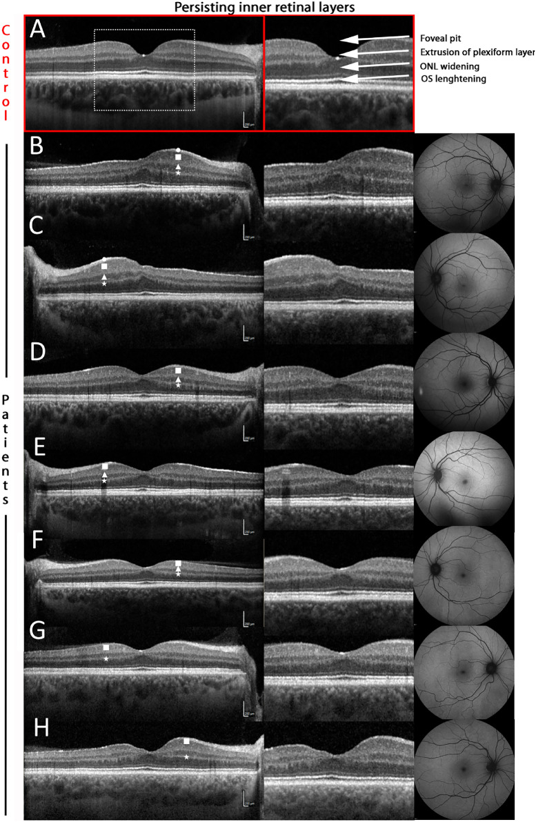
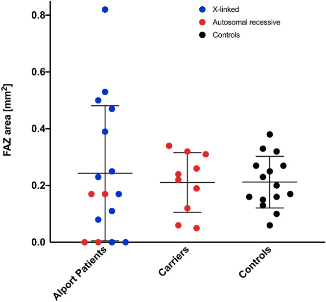
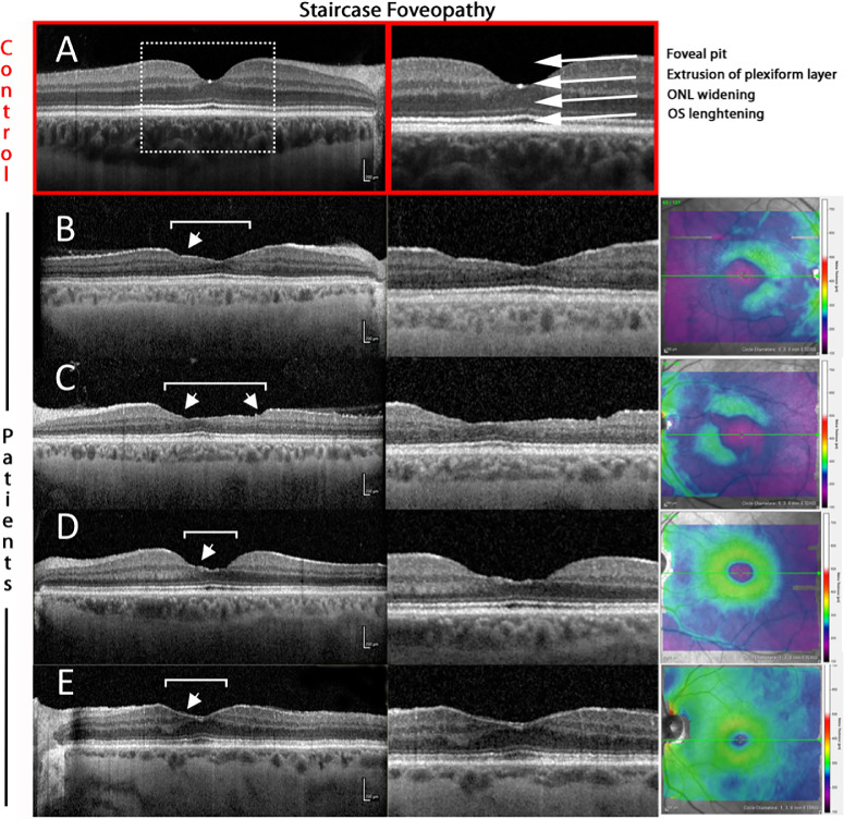
Foveal hypoplasia was found in 10 eyes from seven patients with either genotype. In contrast, a staircase foveopathy was found in seven eyes of four X-linked AS patients. The average FAZ area did not differ significantly between AS patients and control subjects (mean ± SD 0.24 ± 0.24 mm2 vs. 0.21 ± 0.09 mm2; P = 0.64). Five eyes showed absence or severe anomalies of the FAZ with crossing macular capillaries that was linked to the degree of foveal hypoplasia on OCT images leading to a significant inverse correlation of FAZ area and foveal thickness (r = -0.88; P < 0.001). In contrary, female patients with X-linked mutations exhibited a significantly greater FAZ area (0.48 ± 0.30 mm2 vs. 0.21 ± 0.09 mm2; P = 0.007), in line with OCT findings of a staircase foveopathy.

CONCLUSIONS

The foveal phenotypic spectrum in AS ranges from foveal hypoplasia and absence of a FAZ to staircase foveopathy with an enlarged FAZ. Because the development of the FAZ and foveal pit are closely related, these findings suggest an important role for collagen type IV in foveal development and maturation.

摘要

目的

研究 Alport 综合征(AS)患者黄斑中心凹小凹和无血管区(FAZ)的特征。AS 是一种罕见的单基因疾病,由编码 IV 型胶原的基因突变引起。

方法

使用光学相干断层扫描(OCT)和 OCT 血管造影(OCT-A)检查 9 例 AS 患者的 28 只眼、5 名常染色体隐性携带者和 15 名年龄匹配的健康对照者的 15 只眼。将黄斑中心凹形态和 FAZ 测量值(包括 FAZ 面积、圆度和中央 1°和 3°的血管密度)进行相关性分析。

结果

7 名患者的 10 只眼发现黄斑中心凹发育不良,而 4 名 X 连锁 AS 患者的 7 只眼发现阶梯状黄斑病变。AS 患者和对照组的 FAZ 面积无显著差异(平均 ± SD 0.24 ± 0.24mm2与 0.21 ± 0.09mm2;P = 0.64)。5 只眼的 FAZ 缺失或严重异常,伴有黄斑毛细血管交叉,与 OCT 图像上黄斑中心凹发育不良的程度相关,导致 FAZ 面积与黄斑中心凹厚度呈显著负相关(r = -0.88;P < 0.001)。相反,X 连锁突变的女性患者 FAZ 面积显著增大(0.48 ± 0.30mm2与 0.21 ± 0.09mm2;P = 0.007),与阶梯状黄斑病变的 OCT 发现一致。

结论

AS 的黄斑表型谱从黄斑中心凹发育不良和 FAZ 缺失到阶梯状黄斑病变伴 FAZ 增大。由于 FAZ 和黄斑中心凹小凹的发育密切相关,这些发现表明 IV 型胶原在黄斑中心凹发育和成熟中起着重要作用。

https://cdn.ncbi.nlm.nih.gov/pmc/blobs/b44f/7324255/db2198300be5/iovs-61-2-5-f007.jpg
https://cdn.ncbi.nlm.nih.gov/pmc/blobs/b44f/7324255/da8d55659043/iovs-61-2-5-f001.jpg
https://cdn.ncbi.nlm.nih.gov/pmc/blobs/b44f/7324255/59b4e0385a94/iovs-61-2-5-f002.jpg
https://cdn.ncbi.nlm.nih.gov/pmc/blobs/b44f/7324255/7dd7af6c6bb4/iovs-61-2-5-f003.jpg
https://cdn.ncbi.nlm.nih.gov/pmc/blobs/b44f/7324255/1961f769fff8/iovs-61-2-5-f004.jpg
https://cdn.ncbi.nlm.nih.gov/pmc/blobs/b44f/7324255/52ca09d227a9/iovs-61-2-5-f005.jpg
https://cdn.ncbi.nlm.nih.gov/pmc/blobs/b44f/7324255/d6f91b9e042e/iovs-61-2-5-f006.jpg
https://cdn.ncbi.nlm.nih.gov/pmc/blobs/b44f/7324255/db2198300be5/iovs-61-2-5-f007.jpg

相似文献

1
Phenotypic Spectrum of the Foveal Configuration and Foveal Avascular Zone in Patients With Alport Syndrome.Alport 综合征患者的黄斑形态与黄斑无血管区的表型谱。
Invest Ophthalmol Vis Sci. 2020 Feb 7;61(2):5. doi: 10.1167/iovs.61.2.5.
2
Visual Acuity and Foveal Structure in Eyes with Fragmented Foveal Avascular Zones.伴有部分性黄斑区无血管区的眼的视力和中心凹结构。
Ophthalmol Retina. 2020 May;4(5):535-544. doi: 10.1016/j.oret.2019.11.014. Epub 2019 Nov 22.
3
Swept-Source OCT Angiography of the Anomalous Foveal Avascular Zone.异常黄斑无血管区的扫频源光学相干断层扫描血管造影
Ophthalmic Surg Lasers Imaging Retina. 2018 Nov 1;49(11):852-858. doi: 10.3928/23258160-20181101-06.
4
[Correlation of capillary plexus with visual acuity in idiopathic macular epiretinal membrane eyes using optical coherence tomography angiography].[利用光学相干断层扫描血管造影术研究特发性黄斑视网膜前膜眼中毛细血管丛与视力的相关性]
Zhonghua Yan Ke Za Zhi. 2019 Oct 11;55(10):757-762. doi: 10.3760/cma.j.issn.0412-4081.2019.10.006.
5
OPTICAL COHERENCE TOMOGRAPHY ANGIOGRAPHY OF THE FOVEA IN CHILDREN BORN PRETERM.早产儿黄斑区的光学相干断层扫描血管造影
Retina. 2017 Dec;37(12):2289-2294. doi: 10.1097/IAE.0000000000001471.
6
Foveal ganglion cell layer damage in ischemic diabetic maculopathy: correlation of optical coherence tomographic and anatomic changes.缺血性糖尿病性黄斑病变中的黄斑神经节细胞层损伤:光学相干断层扫描与解剖学变化的相关性
Ophthalmology. 2009 Oct;116(10):1949-59.e8. doi: 10.1016/j.ophtha.2009.06.066. Epub 2009 Aug 21.
7
Foveal Hypoplasia in Patients with Stickler Syndrome.斯特奇-韦伯综合征患者的中心凹发育不良。
Ophthalmology. 2017 Jun;124(6):896-902. doi: 10.1016/j.ophtha.2017.01.046. Epub 2017 Mar 7.
8
Optical Coherence Tomography Angiography in Familial Exudative Vitreoretinopathy: Clinical Features and Phenotype-Genotype Correlation.光学相干断层扫描血管造影在家族性渗出性玻璃体视网膜病变中的应用:临床特征及表型-基因型相关性。
Invest Ophthalmol Vis Sci. 2018 Dec 3;59(15):5726-5734. doi: 10.1167/iovs.18-25377.
9
Foveal microvascular anomalies on optical coherence tomography angiography and the correlation with foveal thickness and visual acuity in retinopathy of prematurity.早产儿视网膜病变中光学相干断层扫描血管造影的黄斑微血管异常及其与黄斑厚度和视力的相关性
Graefes Arch Clin Exp Ophthalmol. 2019 Jan;257(1):23-30. doi: 10.1007/s00417-018-4162-y. Epub 2018 Oct 25.
10
Alterations of the Foveal Avascular Zone Measured by Optical Coherence Tomography Angiography in Glaucoma Patients With Central Visual Field Defects.通过光学相干断层扫描血管造影术测量的青光眼中央视野缺损患者黄斑无血管区的改变
Invest Ophthalmol Vis Sci. 2017 Mar 1;58(3):1637-1645. doi: 10.1167/iovs.16-21079.

引用本文的文献

1
Stair-Case/Honeycomb Maculopathy in Alport Syndrome: A Case Report.Alport综合征中的阶梯状/蜂窝状黄斑病变:一例报告
Case Rep Ophthalmol. 2025 Jun 24;16(1):496-502. doi: 10.1159/000546567. eCollection 2025 Jan-Dec.
2
Diagnosis, management and treatment of the Alport syndrome - 2024 guideline on behalf of ERKNet, ERA and ESPN.奥尔波特综合征的诊断、管理与治疗——2024年代表ERKNet、ERA和ESPN发布的指南
Nephrol Dial Transplant. 2025 May 30;40(6):1091-1106. doi: 10.1093/ndt/gfae265.
3
Electroretinographic abnormalities in Alport syndrome with a novel COL4A5 truncated variant (p.Try20GlyfsTer19).

本文引用的文献

1
Features of Autosomal Recessive Alport Syndrome: A Systematic Review.常染色体隐性遗传性奥尔波特综合征的特征:一项系统综述。
J Clin Med. 2019 Feb 3;8(2):178. doi: 10.3390/jcm8020178.
2
Optical Coherence Tomography Angiography in Familial Exudative Vitreoretinopathy: Clinical Features and Phenotype-Genotype Correlation.光学相干断层扫描血管造影在家族性渗出性玻璃体视网膜病变中的应用:临床特征及表型-基因型相关性。
Invest Ophthalmol Vis Sci. 2018 Dec 3;59(15):5726-5734. doi: 10.1167/iovs.18-25377.
3
Should We Diagnose Autosomal Dominant Alport Syndrome When There Is a Pathogenic Heterozygous or Variant?
Alport 综合征中新型 COL4A5 截断变异(p.Try20GlyfsTer19)引起的视网膜电图异常。
Doc Ophthalmol. 2023 Jun;146(3):281-291. doi: 10.1007/s10633-023-09935-w. Epub 2023 May 10.
4
Ocular Manifestations and Potential Treatments of Alport Syndrome: A Systematic Review.奥尔波特综合征的眼部表现及潜在治疗方法:一项系统综述
J Ophthalmol. 2022 Sep 8;2022:9250367. doi: 10.1155/2022/9250367. eCollection 2022.
5
Foveal Curvature and Its Associations in UK Biobank Participants.英国生物库参与者的中央凹曲率及其相关性。
Invest Ophthalmol Vis Sci. 2022 Jul 8;63(8):26. doi: 10.1167/iovs.63.8.26.
6
Characterization of Choriocapillaris and Choroidal Abnormalities in Alport Syndrome.Alport 综合征的脉络膜毛细血管和脉络膜异常特征。
Transl Vis Sci Technol. 2022 Mar 2;11(3):23. doi: 10.1167/tvst.11.3.23.
当存在致病性杂合子或变异时,我们是否应诊断常染色体显性遗传性阿尔波特综合征?
Kidney Int Rep. 2018 Aug 22;3(6):1239-1241. doi: 10.1016/j.ekir.2018.08.002. eCollection 2018 Nov.
4
Comparison of methods to quantify macular and peripapillary vessel density in optical coherence tomography angiography.比较光学相干断层扫描血管造影术定量黄斑和视盘周围血管密度的方法。
PLoS One. 2018 Oct 18;13(10):e0205773. doi: 10.1371/journal.pone.0205773. eCollection 2018.
5
INNER MACULAR CHANGES AFTER VITRECTOMY WITH INTERNAL LIMITING MEMBRANE PEELING FOR RHEGMATOGENOUS RETINAL DETACHMENT: Similarity With Alport Syndrome.孔源性视网膜脱离行玻璃体切除联合内界膜剥除术后的黄斑内层改变:与 Alport 综合征的相似性。
Retina. 2019 Dec;39(12):2332-2340. doi: 10.1097/IAE.0000000000002310.
6
A review of clinical characteristics and genetic backgrounds in Alport syndrome.奥尔波特综合征的临床特征与遗传背景综述。
Clin Exp Nephrol. 2019 Feb;23(2):158-168. doi: 10.1007/s10157-018-1629-4. Epub 2018 Aug 20.
7
Detection of Choriocapillaris Loss in Alport Syndrome With Swept-Source OCT Angiography.使用扫频光学相干断层扫描血管造影术检测Alport综合征中的脉络膜毛细血管层缺失
Ophthalmic Surg Lasers Imaging Retina. 2018 Feb 1;49(2):138-141. doi: 10.3928/23258160-20180129-10.
8
Foveal Hypoplasia in Patients with Stickler Syndrome.斯特奇-韦伯综合征患者的中心凹发育不良。
Ophthalmology. 2017 Jun;124(6):896-902. doi: 10.1016/j.ophtha.2017.01.046. Epub 2017 Mar 7.
9
Optical coherence tomography angiography of foveal hypoplasia.黄斑发育不全的光学相干断层扫描血管造影
Br J Ophthalmol. 2017 Jul;101(7):985-988. doi: 10.1136/bjophthalmol-2016-309200. Epub 2016 Nov 29.
10
VEGF-A/Notch-Induced Podosomes Proteolyse Basement Membrane Collagen-IV during Retinal Sprouting Angiogenesis.VEGF-A/Notch诱导的足体在视网膜新生血管形成过程中降解基底膜胶原蛋白IV
Cell Rep. 2016 Oct 4;17(2):484-500. doi: 10.1016/j.celrep.2016.09.016.