• 文献检索
  • 文档翻译
  • 深度研究
  • 学术资讯
  • Suppr Zotero 插件Zotero 插件
  • 邀请有礼
  • 套餐&价格
  • 历史记录
应用&插件
Suppr Zotero 插件Zotero 插件浏览器插件Mac 客户端Windows 客户端微信小程序
定价
高级版会员购买积分包购买API积分包
服务
文献检索文档翻译深度研究API 文档MCP 服务
关于我们
关于 Suppr公司介绍联系我们用户协议隐私条款
关注我们

Suppr 超能文献

核心技术专利:CN118964589B侵权必究
粤ICP备2023148730 号-1Suppr @ 2026

文献检索

告别复杂PubMed语法,用中文像聊天一样搜索,搜遍4000万医学文献。AI智能推荐,让科研检索更轻松。

立即免费搜索

文件翻译

保留排版,准确专业,支持PDF/Word/PPT等文件格式,支持 12+语言互译。

免费翻译文档

深度研究

AI帮你快速写综述,25分钟生成高质量综述,智能提取关键信息,辅助科研写作。

立即免费体验

蛋白激酶 D 家族激酶在破骨细胞分化的多个阶段的调节作用。

Regulation of Osteoclast Differentiation at Multiple Stages by Protein Kinase D Family Kinases.

机构信息

Department of Diagnostic and Biological Sciences, University of Minnesota School of Dentistry, Minneapolis, MN 55455, USA.

Clinical and Translational Science Institute, University of Minnesota, Minneapolis, MN 55455, USA.

出版信息

Int J Mol Sci. 2020 Feb 5;21(3):1056. doi: 10.3390/ijms21031056.

DOI:10.3390/ijms21031056
PMID:32033440
原文链接:https://pmc.ncbi.nlm.nih.gov/articles/PMC7036879/
Abstract

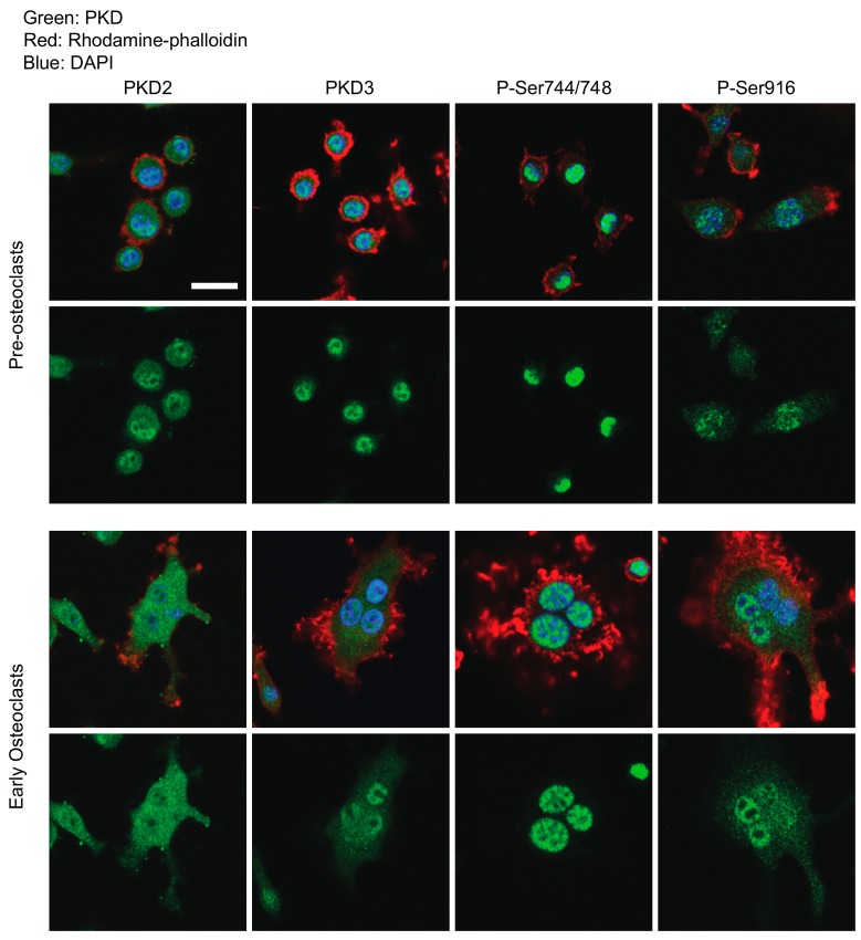
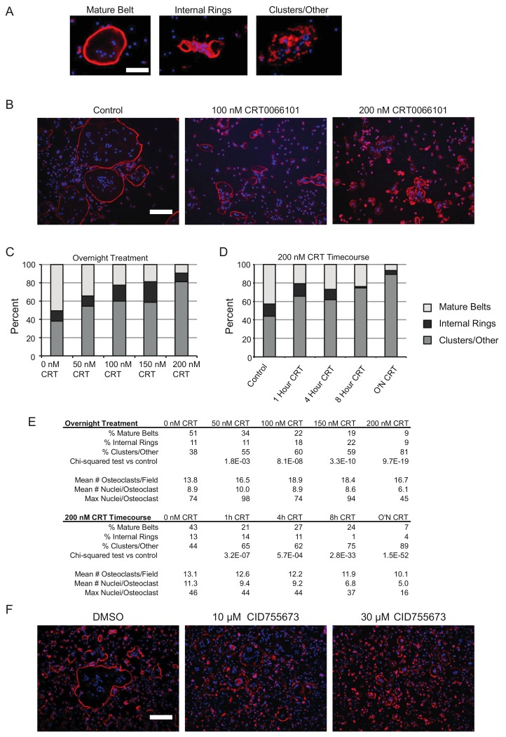
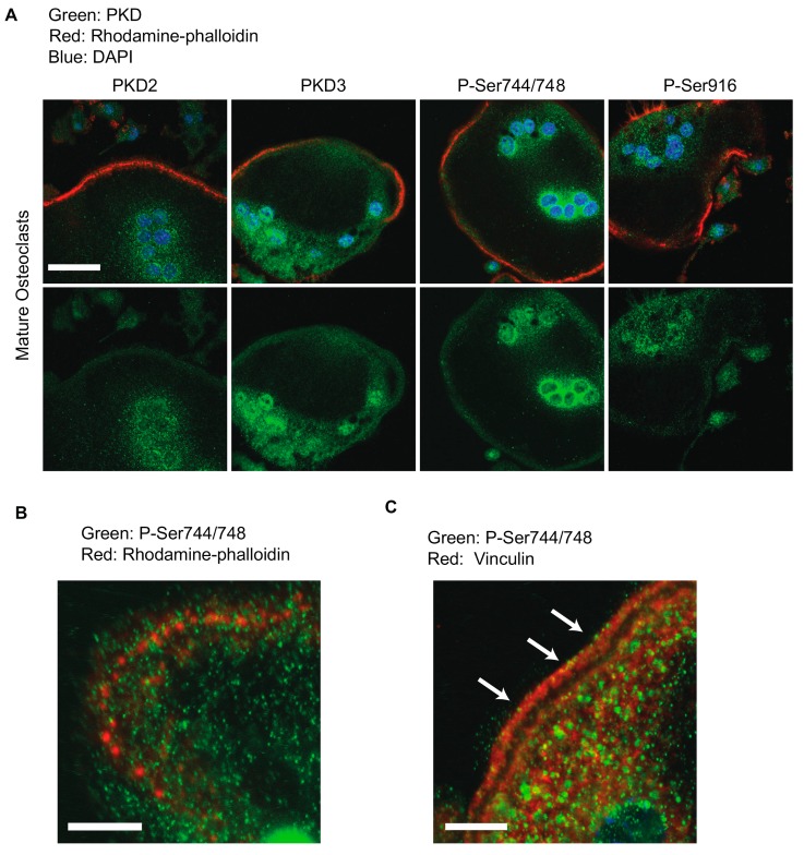
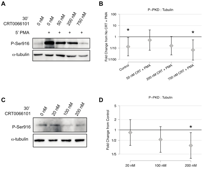
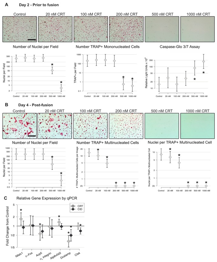
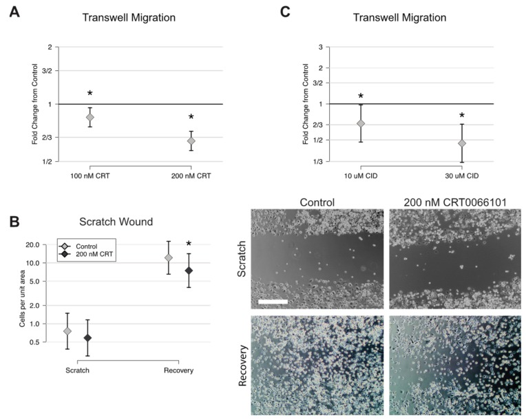
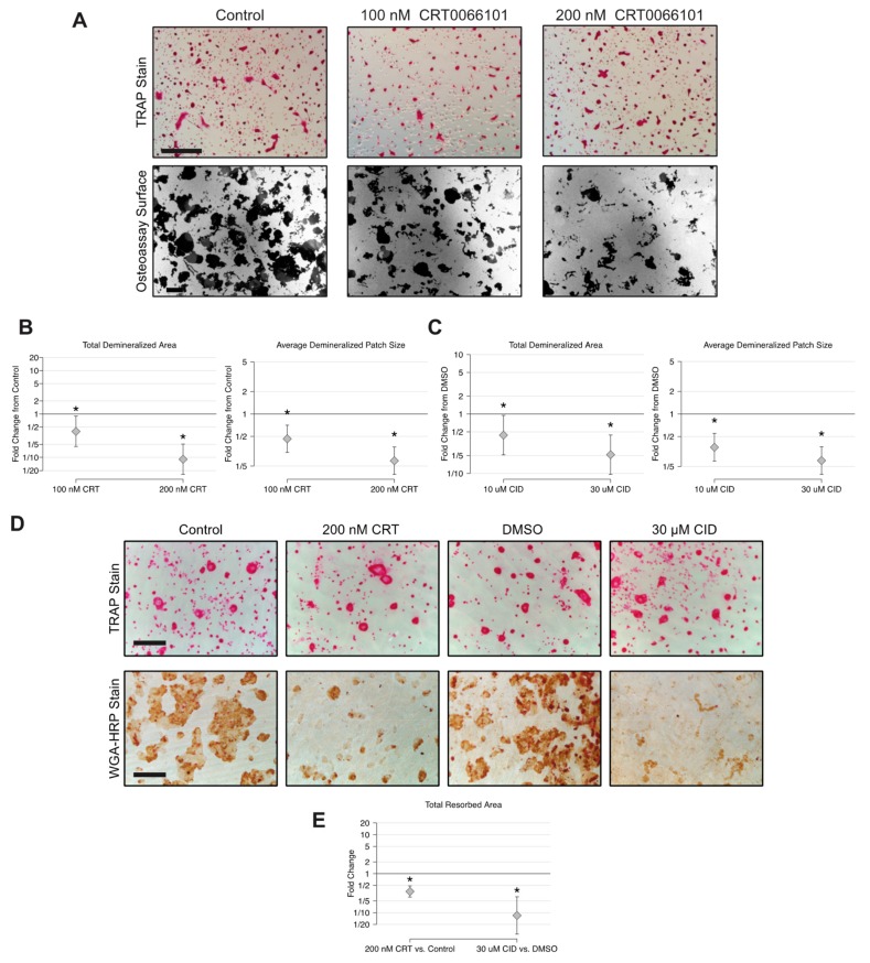
Balanced osteoclast and osteoblast activity is necessary for skeletal health, whereas unbalanced osteoclast activity causes bone loss in many skeletal conditions. A better understanding of pathways that regulate osteoclast differentiation and activity is necessary for the development of new therapies to better manage bone resorption. The roles of Protein Kinase D (PKD) family of serine/threonine kinases in osteoclasts have not been well characterized. In this study we use immunofluorescence analysis to reveal that PKD2 and PKD3, the isoforms expressed in osteoclasts, are found in the nucleus and cytoplasm, the mitotic spindle and midbody, and in association with the actin belt. We show that PKD inhibitors CRT0066101 and CID755673 inhibit several distinct aspects of osteoclast formation. Treating bone marrow macrophages with lower doses of the PKD inhibitors had little effect on M-CSF + RANKL-dependent induction into committed osteoclast precursors, but inhibited their motility and subsequent differentiation into multinucleated mature osteoclasts, whereas higher doses of the PKD inhibitors induced apoptosis of the preosteoclasts. Treating post-fusion multinucleated osteoclasts with the inhibitors disrupted the osteoclast actin belts and impaired their resorptive activity. In conclusion, these data implicate PKD kinases as positive regulators of osteoclasts, which are essential for multiple distinct processes throughout their formation and function.

摘要

成骨细胞和破骨细胞活性的平衡对于骨骼健康是必要的,而破骨细胞活性的失衡会导致许多骨骼疾病中的骨质流失。为了开发新的治疗方法来更好地控制骨质吸收,需要更好地了解调节破骨细胞分化和活性的途径。蛋白激酶 D (PKD) 家族丝氨酸/苏氨酸激酶在破骨细胞中的作用尚未得到很好的描述。在这项研究中,我们使用免疫荧光分析揭示了在破骨细胞中表达的 PKD2 和 PKD3,它们存在于细胞核和细胞质、有丝分裂纺锤体和中体中,并与肌动蛋白带相关联。我们表明,PKD 抑制剂 CRT0066101 和 CID755673 抑制破骨细胞形成的几个不同方面。用较低剂量的 PKD 抑制剂处理骨髓巨噬细胞对 M-CSF + RANKL 依赖性诱导成 committed 破骨细胞前体的作用很小,但抑制了它们的迁移和随后分化为多核成熟破骨细胞,而较高剂量的 PKD 抑制剂则诱导前破骨细胞凋亡。用抑制剂处理融合后的多核破骨细胞会破坏破骨细胞的肌动蛋白带并损害其吸收活性。总之,这些数据表明 PKD 激酶是破骨细胞的正调控因子,对于破骨细胞形成和功能的多个不同过程都是必不可少的。

https://cdn.ncbi.nlm.nih.gov/pmc/blobs/2a7c/7036879/81505a42c574/ijms-21-01056-g007.jpg
https://cdn.ncbi.nlm.nih.gov/pmc/blobs/2a7c/7036879/d0be64a73a96/ijms-21-01056-g001.jpg
https://cdn.ncbi.nlm.nih.gov/pmc/blobs/2a7c/7036879/7217055ccf14/ijms-21-01056-g002.jpg
https://cdn.ncbi.nlm.nih.gov/pmc/blobs/2a7c/7036879/40ea3ec422bb/ijms-21-01056-g003.jpg
https://cdn.ncbi.nlm.nih.gov/pmc/blobs/2a7c/7036879/1ed4cc44bd6b/ijms-21-01056-g004.jpg
https://cdn.ncbi.nlm.nih.gov/pmc/blobs/2a7c/7036879/b7bd4cdc4fb7/ijms-21-01056-g005.jpg
https://cdn.ncbi.nlm.nih.gov/pmc/blobs/2a7c/7036879/e6e67605bf33/ijms-21-01056-g006.jpg
https://cdn.ncbi.nlm.nih.gov/pmc/blobs/2a7c/7036879/81505a42c574/ijms-21-01056-g007.jpg

相似文献

1
Regulation of Osteoclast Differentiation at Multiple Stages by Protein Kinase D Family Kinases.蛋白激酶 D 家族激酶在破骨细胞分化的多个阶段的调节作用。
Int J Mol Sci. 2020 Feb 5;21(3):1056. doi: 10.3390/ijms21031056.
2
Osteoblasts/stromal cells stimulate osteoclast activation through expression of osteoclast differentiation factor/RANKL but not macrophage colony-stimulating factor: receptor activator of NF-kappa B ligand.成骨细胞/基质细胞通过表达破骨细胞分化因子/核因子κB受体活化因子配体(RANKL)而非巨噬细胞集落刺激因子来刺激破骨细胞活化:核因子κB受体活化因子配体。
Bone. 1999 Nov;25(5):517-23. doi: 10.1016/s8756-3282(99)00210-0.
3
Saurolactam inhibits osteoclast differentiation and stimulates apoptosis of mature osteoclasts.蜥内酰胺抑制破骨细胞分化并刺激成熟破骨细胞凋亡。
J Cell Physiol. 2009 Dec;221(3):618-28. doi: 10.1002/jcp.21892.
4
Protein kinase D promotes in vitro osteoclast differentiation and fusion.蛋白激酶 D 促进体外破骨细胞分化和融合。
J Biol Chem. 2013 Apr 5;288(14):9826-9834. doi: 10.1074/jbc.M112.444133. Epub 2013 Feb 21.
5
RANKL coordinates cell cycle withdrawal and differentiation in osteoclasts through the cyclin-dependent kinase inhibitors p27KIP1 and p21CIP1.核因子κB受体活化因子配体(RANKL)通过细胞周期蛋白依赖性激酶抑制剂p27KIP1和p21CIP1来协调破骨细胞的细胞周期停滞和分化。
J Bone Miner Res. 2004 Aug;19(8):1339-48. doi: 10.1359/JBMR.040321. Epub 2004 Mar 29.
6
Chloroform extract of deer antler inhibits osteoclast differentiation and bone resorption.鹿茸的氯仿提取物可抑制破骨细胞分化和骨吸收。
J Ethnopharmacol. 2007 Sep 5;113(2):191-8. doi: 10.1016/j.jep.2007.04.020. Epub 2007 May 16.
7
BSP and RANKL induce osteoclastogenesis and bone resorption synergistically.骨唾液蛋白(BSP)和核因子κB受体活化因子配体(RANKL)协同诱导破骨细胞生成和骨吸收。
J Bone Miner Res. 2005 Sep;20(9):1669-79. doi: 10.1359/JBMR.050511. Epub 2005 May 16.
8
Bone morphogenetic protein 2 stimulates osteoclast differentiation and survival supported by receptor activator of nuclear factor-kappaB ligand.骨形态发生蛋白2在核因子κB受体激活剂配体的支持下刺激破骨细胞分化和存活。
Endocrinology. 2001 Aug;142(8):3656-62. doi: 10.1210/endo.142.8.8300.
9
Tumor necrosis factor alpha stimulates osteoclast differentiation by a mechanism independent of the ODF/RANKL-RANK interaction.肿瘤坏死因子α通过一种独立于骨桥蛋白/核因子κB受体活化因子配体-核因子κB受体活化因子相互作用的机制刺激破骨细胞分化。
J Exp Med. 2000 Jan 17;191(2):275-86. doi: 10.1084/jem.191.2.275.
10
Inhibition of osteoclast differentiation and bone resorption by a novel lysophosphatidylcholine derivative, SCOH.新型溶血磷脂酰胆碱衍生物SCOH对破骨细胞分化和骨吸收的抑制作用
Biochem Pharmacol. 2004 Apr 1;67(7):1239-48. doi: 10.1016/j.bcp.2003.10.032.

引用本文的文献

1
CircPRKD3/miR-6783-3p responds to mechanical force to facilitate the osteogenesis of stretched periodontal ligament stem cells.环状PRKD3/miR-6783-3p响应机械力以促进拉伸的牙周膜干细胞的成骨作用。
J Orthop Surg Res. 2024 Apr 22;19(1):257. doi: 10.1186/s13018-024-04727-7.
2
Effect of protein kinase D inhibitor CRT0066101 on the cell migration of salivary adenoid cystic carcinoma.蛋白激酶D抑制剂CRT0066101对涎腺腺样囊性癌细胞迁移的影响
Hua Xi Kou Qiang Yi Xue Za Zhi. 2022 May 25;40(3):320-327. doi: 10.7518/hxkq.2022.03.012.
3
TRAF-STOP alleviates osteoclastogenesis in periodontitis.

本文引用的文献

1
Protein Kinase D2 Modulates Cell Cycle By Stabilizing Aurora A Kinase at Centrosomes.蛋白激酶 D2 通过稳定中心体处的 Aurora A 激酶来调节细胞周期。
Mol Cancer Res. 2018 Nov;16(11):1785-1797. doi: 10.1158/1541-7786.MCR-18-0641. Epub 2018 Jul 17.
2
Common signalling pathways in macrophage and osteoclast multinucleation.巨噬细胞和破骨细胞多核化的常见信号通路。
J Cell Sci. 2018 Jun 5;131(11):jcs216267. doi: 10.1242/jcs.216267.
3
The Sealing Zone in Osteoclasts: A Self-Organized Structure on the Bone.破骨细胞中的密封区:骨表面的自组织结构
TRAF-STOP可减轻牙周炎中的破骨细胞生成。
Front Pharmacol. 2023 May 15;14:1119847. doi: 10.3389/fphar.2023.1119847. eCollection 2023.
4
Histone deacetylase 5 is a phosphorylation substrate of protein kinase D in osteoclasts.组蛋白去乙酰化酶5是破骨细胞中蛋白激酶D的磷酸化底物。
Bone. 2022 Jun;159:116393. doi: 10.1016/j.bone.2022.116393. Epub 2022 Mar 19.
5
Small-Molecule Inhibitor Targeting Protein Kinase D: A Potential Therapeutic Strategy.靶向蛋白激酶D的小分子抑制剂:一种潜在的治疗策略。
Front Oncol. 2021 Jun 24;11:680221. doi: 10.3389/fonc.2021.680221. eCollection 2021.
Int J Mol Sci. 2018 Mar 26;19(4):984. doi: 10.3390/ijms19040984.
4
Novel Small Molecule Inhibitors of Protein Kinase D Suppress NF-kappaB Activation and Attenuate the Severity of Rat Cerulein Pancreatitis.蛋白激酶D的新型小分子抑制剂可抑制核因子κB激活并减轻大鼠雨蛙肽诱导的胰腺炎严重程度。
Front Physiol. 2017 Dec 7;8:1014. doi: 10.3389/fphys.2017.01014. eCollection 2017.
5
Antiresorptives: Safety Concerns-Clinical Perspective.抗吸收剂:安全问题——临床视角
Toxicol Pathol. 2017 Oct;45(7):859-863. doi: 10.1177/0192623317737066. Epub 2017 Oct 24.
6
Protein kinase D signaling in cancer: A friend or foe?蛋白激酶 D 信号通路与癌症:是敌是友?
Biochim Biophys Acta Rev Cancer. 2017 Aug;1868(1):283-294. doi: 10.1016/j.bbcan.2017.05.008. Epub 2017 May 31.
7
Insulin Receptor and GPCR Crosstalk Stimulates YAP via PI3K and PKD in Pancreatic Cancer Cells.胰岛素受体与G蛋白偶联受体的相互作用通过PI3K和PKD在胰腺癌细胞中刺激YAP
Mol Cancer Res. 2017 Jul;15(7):929-941. doi: 10.1158/1541-7786.MCR-17-0023. Epub 2017 Mar 30.
8
A Conditional Knockout Mouse Model Reveals a Critical Role of PKD1 in Osteoblast Differentiation and Bone Development.条件性基因敲除小鼠模型揭示 PKD1 在成骨细胞分化和骨发育中的关键作用。
Sci Rep. 2017 Jan 13;7:40505. doi: 10.1038/srep40505.
9
Bradykinin stimulates protein kinase D-mediated colonic myofibroblast migration via cyclooxygenase-2 and heat shock protein 27.缓激肽通过环氧化酶-2和热休克蛋白27刺激蛋白激酶D介导的结肠肌成纤维细胞迁移。
J Surg Res. 2017 Mar;209:191-198. doi: 10.1016/j.jss.2016.10.014. Epub 2016 Oct 20.
10
Protein Kinase D and Gβγ Subunits Mediate Agonist-evoked Translocation of Protease-activated Receptor-2 from the Golgi Apparatus to the Plasma Membrane.蛋白激酶D和Gβγ亚基介导激动剂诱发的蛋白酶激活受体-2从高尔基体向质膜的转运。
J Biol Chem. 2016 May 20;291(21):11285-99. doi: 10.1074/jbc.M115.710681. Epub 2016 Mar 30.