Department of Experimental Medical Science, Division of Neuroscience, Glycobiology Group, Lund University, 221 00 Lund, Sweden.
Glycobiology. 2020 Jul 16;30(8):539-549. doi: 10.1093/glycob/cwaa011.
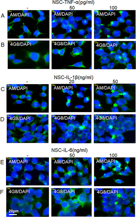
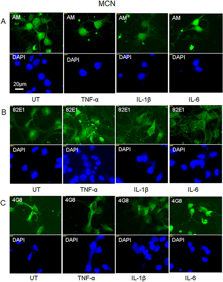
Proinflammatory cytokines stimulate expression of β-secretase, which increases processing of amyloid precursor protein (APP), ultimately leading to the deposition of amyloid beta (Aβ). The N-terminal domain of β-cleaved APP supports Cu/NO-dependent release of heparan sulfate (HS) from the glypican-1 (Gpc-1) proteoglycan. HS is an inhibitor of β-secretase, thereby constituting a regulatory, negative feedback loop. Here, we have investigated the effect of the proinflammatory cytokines TNF-α, IL-1β and IL-6 on the interplay between APP processing and release of HS from Gpc-1 in neuronal cells. We have used deconvolution immunofluorescence microscopy and sodium dodecyl sulfate polyacrylamide gel electrophoresis (SDS-PAGE) and a panel of monoclonal/polyclonal antibodies recognizing the released HS, the N-terminus of Aβ, Aβ, the C-terminus of APP and the autophagosome marker LC3 as well as the chemical lysosome marker LysoTrackerRed (LTR). We repeatedly found that N2a neuroblastoma cells and human neural stem cells grown in the presence of the cytokines developed large cytoplasmic clusters, which stained positive for HS, the N-terminus of Aβ, Aβ, the C-terminus of APP, LC3 and LTR, indicating accumulation of HS and APP/APP degradation products in enlarged autophagosomes/lysosomes. The SDS-PAGE of immunoisolates obtained from TNF-α-treated N2a cells by using anti-C-terminus of APP revealed the presence of SDS-stable complexes between HS and the C-terminal fragment of β-cleaved APP (βCTF) migrating in the range 10-18 kDa. Clustered accumulation of βCTF disappeared when HS release was prevented and slightly enhanced when HS release was increased. Hence, when proinflammatory cytokines induce increased processing of APP, inhibition of β-secretase by HS is insufficient, which may lead to the impaired autophagosomal degradation.
促炎细胞因子刺激β-分泌酶的表达,增加淀粉样前体蛋白 (APP) 的加工,最终导致淀粉样β (Aβ) 的沉积。β-切割 APP 的 N 端结构域支持糖蛋白聚糖-1 (Gpc-1) 蛋白聚糖中肝素硫酸酯 (HS) 的 Cu/NO 依赖性释放。HS 是 β-分泌酶的抑制剂,从而构成调节性负反馈环。在这里,我们研究了促炎细胞因子 TNF-α、IL-1β 和 IL-6 对神经元细胞中 APP 加工和 Gpc-1 中 HS 释放之间相互作用的影响。我们使用反卷积免疫荧光显微镜和十二烷基硫酸钠聚丙烯酰胺凝胶电泳 (SDS-PAGE) 以及一组单克隆/多克隆抗体来识别释放的 HS、Aβ 的 N 端、Aβ、APP 的 C 端和自噬体标记物 LC3 以及化学溶酶体标记物 LysoTrackerRed (LTR)。我们反复发现,在细胞因子存在下生长的 N2a 神经母细胞瘤细胞和人神经干细胞会形成大的细胞质簇,这些细胞质簇对 HS、Aβ 的 N 端、Aβ、APP 的 C 端、LC3 和 LTR 呈阳性染色,表明 HS 和 APP/APP 降解产物在扩大的自噬体/溶酶体中积累。用抗 APP C 端从 TNF-α 处理的 N2a 细胞中获得的免疫分离物的 SDS-PAGE 显示,HS 与 β 切割 APP 的 C 端片段 (βCTF) 之间存在 SDS 稳定的复合物,其迁移范围为 10-18 kDa。当阻止 HS 释放时,簇状聚集的 βCTF 消失,当 HS 释放增加时,βCTF 聚集略有增加。因此,当促炎细胞因子诱导 APP 加工增加时,HS 对 β-分泌酶的抑制作用不足,这可能导致自噬体降解受损。