• 文献检索
  • 文档翻译
  • 深度研究
  • 学术资讯
  • Suppr Zotero 插件Zotero 插件
  • 邀请有礼
  • 套餐&价格
  • 历史记录
应用&插件
Suppr Zotero 插件Zotero 插件浏览器插件Mac 客户端Windows 客户端微信小程序
定价
高级版会员购买积分包购买API积分包
服务
文献检索文档翻译深度研究API 文档MCP 服务
关于我们
关于 Suppr公司介绍联系我们用户协议隐私条款
关注我们

Suppr 超能文献

核心技术专利:CN118964589B侵权必究
粤ICP备2023148730 号-1Suppr @ 2026

文献检索

告别复杂PubMed语法,用中文像聊天一样搜索,搜遍4000万医学文献。AI智能推荐,让科研检索更轻松。

立即免费搜索

文件翻译

保留排版,准确专业,支持PDF/Word/PPT等文件格式,支持 12+语言互译。

免费翻译文档

深度研究

AI帮你快速写综述,25分钟生成高质量综述,智能提取关键信息,辅助科研写作。

立即免费体验

利用光片显微镜对拟南芥花中的生殖细胞分化进行成像。

Imaging plant germline differentiation within Arabidopsis flowers by light sheet microscopy.

机构信息

Central European Institute of Technology (CEITEC), Masaryk University, Brno, Czech Republic.

IT4Innovations, VSB-Technical University of Ostrava, Ostrava, Czech Republic.

出版信息

Elife. 2020 Feb 11;9:e52546. doi: 10.7554/eLife.52546.

DOI:10.7554/eLife.52546
PMID:32041682
原文链接:https://pmc.ncbi.nlm.nih.gov/articles/PMC7012603/
Abstract

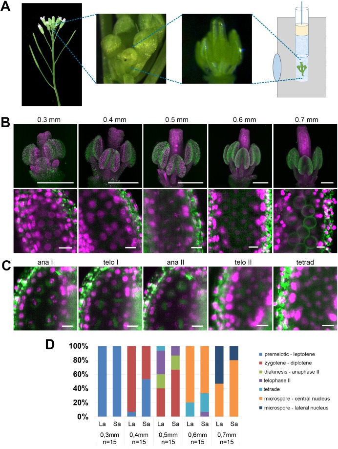
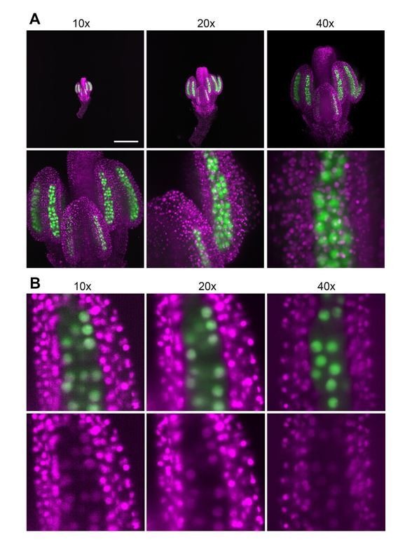
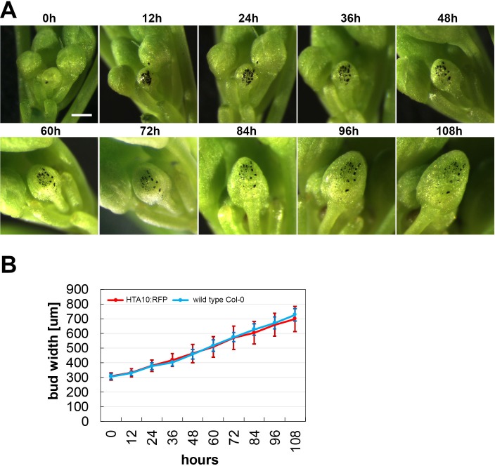
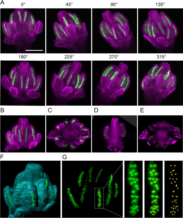
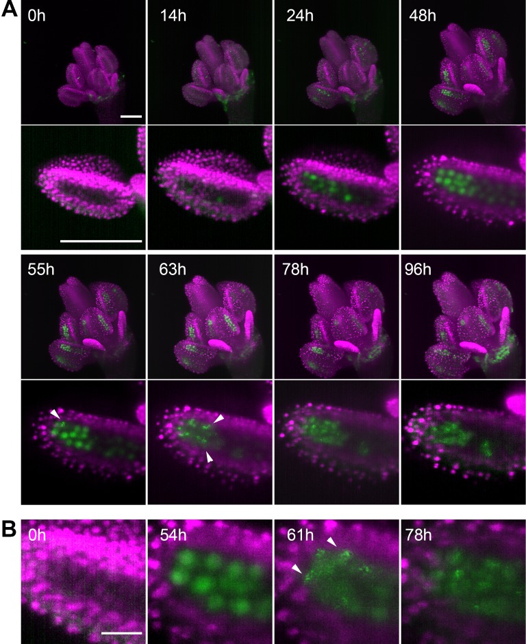
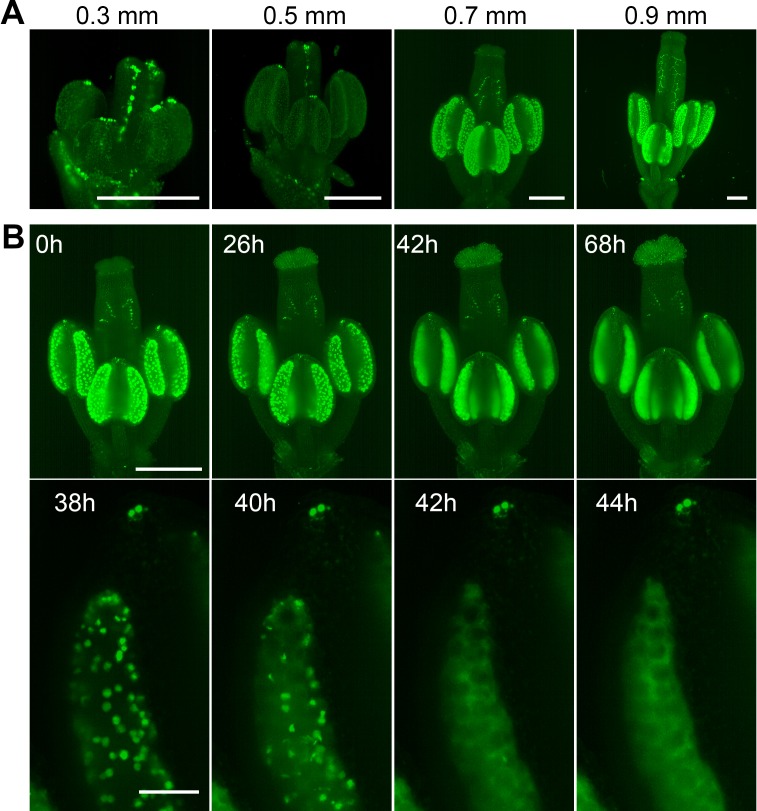
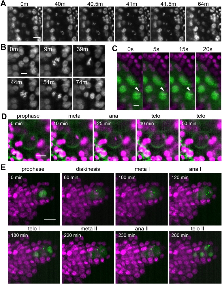
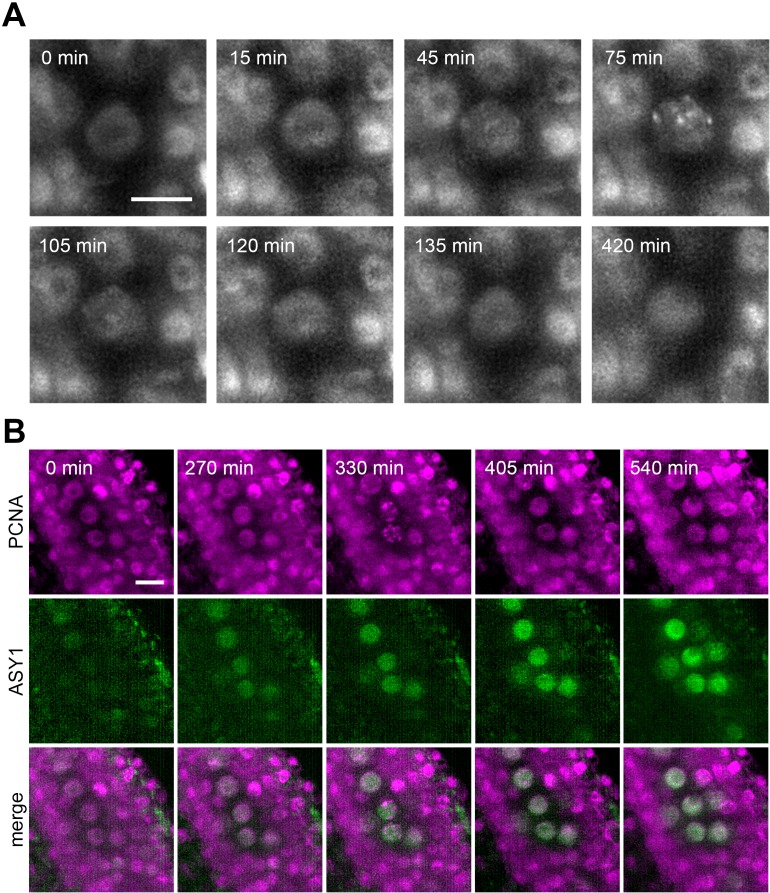
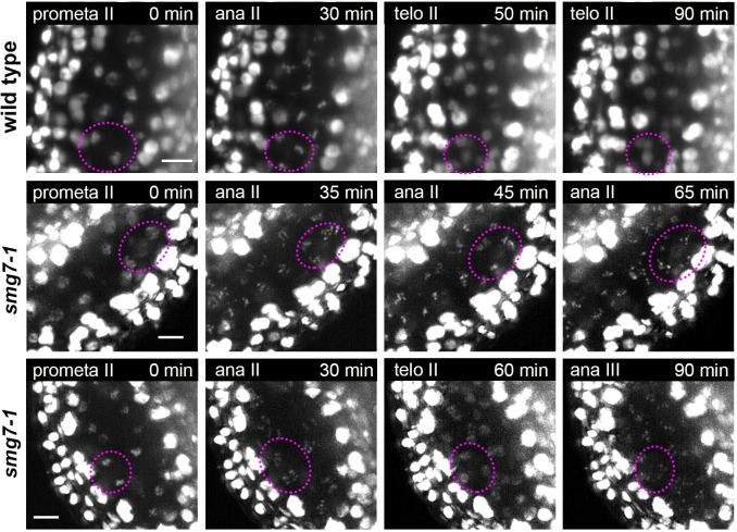
In higher plants, germline differentiation occurs during a relatively short period within developing flowers. Understanding of the mechanisms that govern germline differentiation lags behind other plant developmental processes. This is largely because the germline is restricted to relatively few cells buried deep within floral tissues, which makes them difficult to study. To overcome this limitation, we have developed a methodology for live imaging of the germ cell lineage within floral organs of Arabidopsis using light sheet fluorescence microscopy. We have established reporter lines, cultivation conditions, and imaging protocols for high-resolution microscopy of developing flowers continuously for up to several days. We used multiview imagining to reconstruct a three-dimensional model of a flower at subcellular resolution. We demonstrate the power of this approach by capturing male and female meiosis, asymmetric pollen division, movement of meiotic chromosomes, and unusual restitution mitosis in tapetum cells. This method will enable new avenues of research into plant sexual reproduction.

摘要

在高等植物中,生殖细胞的分化发生在发育中的花朵内相对较短的一段时间内。对控制生殖细胞分化的机制的理解落后于其他植物发育过程。这主要是因为生殖细胞局限于花组织内相对较少的深埋细胞,这使得它们难以研究。为了克服这一限制,我们开发了一种使用光片荧光显微镜对拟南芥花器官中的生殖细胞谱系进行活体成像的方法。我们已经建立了报告基因系、培养条件和成像方案,用于对发育中的花朵进行高分辨率显微镜连续观察长达数天。我们使用多视图成像技术以亚细胞分辨率重建花的三维模型。我们通过捕获雄性和雌性减数分裂、不对称花粉分裂、减数分裂染色体的运动以及绒毡层细胞中的异常有丝后减数分裂,证明了这种方法的有效性。这种方法将为植物有性生殖的研究开辟新途径。

https://cdn.ncbi.nlm.nih.gov/pmc/blobs/b671/7012603/75f29a431680/elife-52546-resp-fig1.jpg
https://cdn.ncbi.nlm.nih.gov/pmc/blobs/b671/7012603/32f99ce2e5a1/elife-52546-fig1.jpg
https://cdn.ncbi.nlm.nih.gov/pmc/blobs/b671/7012603/309d1ce0d809/elife-52546-fig1-figsupp1.jpg
https://cdn.ncbi.nlm.nih.gov/pmc/blobs/b671/7012603/aa0295bde8f4/elife-52546-fig2.jpg
https://cdn.ncbi.nlm.nih.gov/pmc/blobs/b671/7012603/1fe0365595fe/elife-52546-fig3.jpg
https://cdn.ncbi.nlm.nih.gov/pmc/blobs/b671/7012603/86b9328ff5d1/elife-52546-fig3-figsupp1.jpg
https://cdn.ncbi.nlm.nih.gov/pmc/blobs/b671/7012603/43048d99bf26/elife-52546-fig4.jpg
https://cdn.ncbi.nlm.nih.gov/pmc/blobs/b671/7012603/7bf4606befe6/elife-52546-fig5.jpg
https://cdn.ncbi.nlm.nih.gov/pmc/blobs/b671/7012603/3ae82fc3896c/elife-52546-fig6.jpg
https://cdn.ncbi.nlm.nih.gov/pmc/blobs/b671/7012603/37b4f94e35f6/elife-52546-fig7.jpg
https://cdn.ncbi.nlm.nih.gov/pmc/blobs/b671/7012603/75f29a431680/elife-52546-resp-fig1.jpg

相似文献

1
Imaging plant germline differentiation within Arabidopsis flowers by light sheet microscopy.利用光片显微镜对拟南芥花中的生殖细胞分化进行成像。
Elife. 2020 Feb 11;9:e52546. doi: 10.7554/eLife.52546.
2
Germline specification and function in plants.植物的种系发生规范和功能。
Annu Rev Plant Biol. 2011;62:461-84. doi: 10.1146/annurev-arplant-042110-103824.
3
Application of Chemical Inhibitors in Live Cell Imaging of Plant Meiosis Using Light Sheet Fluorescence Microscopy.化学抑制剂在植物减数分裂活细胞光片荧光显微镜成像中的应用
Methods Mol Biol. 2022;2484:93-105. doi: 10.1007/978-1-0716-2253-7_8.
4
Transcriptome analysis of the Arabidopsis megaspore mother cell uncovers the importance of RNA helicases for plant germline development.拟南芥大孢子母细胞转录组分析揭示 RNA 解旋酶在植物生殖细胞发育中的重要性。
PLoS Biol. 2011 Sep;9(9):e1001155. doi: 10.1371/journal.pbio.1001155. Epub 2011 Sep 20.
5
Meiotic and mitotic cell cycle mutants involved in gametophyte development in Arabidopsis.参与拟南芥配子体发育的减数分裂和有丝分裂细胞周期突变体。
Mol Plant. 2008 Jul;1(4):564-74. doi: 10.1093/mp/ssn033.
6
High-resolution live imaging of plant growth in near physiological bright conditions using light sheet fluorescence microscopy.使用光片荧光显微镜在近生理明亮条件下对植物生长进行高分辨率实时成像。
Plant J. 2011 Oct;68(2):377-85. doi: 10.1111/j.1365-313X.2011.04692.x. Epub 2011 Aug 4.
7
A timing mechanism for stem cell maintenance and differentiation in the Arabidopsis floral meristem.拟南芥花分生组织中干细胞维持与分化的定时机制。
Genes Dev. 2009 Aug 1;23(15):1791-804. doi: 10.1101/gad.1800409.
8
Analyzing floral meristem development.分析花分生组织的发育。
Methods Mol Biol. 2010;655:131-42. doi: 10.1007/978-1-60761-765-5_9.
9
High-throughput single-cell transcriptomics reveals the female germline differentiation trajectory in Arabidopsis thaliana.高通量单细胞转录组学揭示了拟南芥雌性生殖细胞的分化轨迹。
Commun Biol. 2021 Oct 1;4(1):1149. doi: 10.1038/s42003-021-02676-z.
10
Graft-transmissible movement of inverted-repeat-induced siRNA signals into flowers.由反向重复序列诱导的小干扰RNA信号通过嫁接在植物体内向花的移动。
Plant J. 2014 Oct;80(1):106-21. doi: 10.1111/tpj.12622. Epub 2014 Aug 14.

引用本文的文献

1
Time-lapse confocal imaging helps to reveal a secret behind gynoecium development.延时共聚焦成像有助于揭示雌蕊发育背后的秘密。
Quant Plant Biol. 2025 Jul 18;6:e18. doi: 10.1017/qpb.2025.10009. eCollection 2025.
2
A Protocol for Live Imaging of Arabidopsis Gynoecium Development Using Confocal Microscopy.一种使用共聚焦显微镜对拟南芥雌蕊发育进行活体成像的方案。
Methods Mol Biol. 2025;2900:351-362. doi: 10.1007/978-1-0716-4398-3_23.
3
Applications of Lightsheet Fluorescence Microscopy by High Numerical Aperture Detection Lens.应用高数值孔径检测透镜的光片荧光显微镜。

本文引用的文献

1
The H3 histone chaperone NASP escorts CenH3 in Arabidopsis.组蛋白 H3 伴侣蛋白 NASP 在拟南芥中护送 CenH3。
Plant J. 2020 Jan;101(1):71-86. doi: 10.1111/tpj.14518. Epub 2019 Oct 14.
2
Live cell imaging of meiosis in .在. 中对减数分裂进行活细胞成像。
Elife. 2019 May 20;8:e42834. doi: 10.7554/eLife.42834.
3
Defining the developmental program leading to meiosis in maize.定义导致玉米减数分裂的发育程序。
J Phys Chem B. 2024 Sep 5;128(35):8273-8289. doi: 10.1021/acs.jpcb.4c01721. Epub 2024 Aug 23.
4
Rapid meiotic prophase chromosome movements in Arabidopsis thaliana are linked to essential reorganization at the nuclear envelope.拟南芥减数分裂前期染色体的快速运动与核膜的必要重排有关。
Nat Commun. 2024 Jul 16;15(1):5964. doi: 10.1038/s41467-024-50169-4.
5
Meiotic double-strand break repair DNA synthesis tracts in Arabidopsis thaliana.拟南芥减数分裂双链断裂修复 DNA 合成片段。
PLoS Genet. 2024 Jul 16;20(7):e1011197. doi: 10.1371/journal.pgen.1011197. eCollection 2024 Jul.
6
Heat stress impairs centromere structure and segregation of meiotic chromosomes in .热应激会损害. 的着丝粒结构和减数分裂染色体的分离。
Elife. 2024 Apr 17;12:RP90253. doi: 10.7554/eLife.90253.
7
Observing ER Dynamics over Long Timescales Using Light Sheet Fluorescence Microscopy.利用光片荧光显微镜观察长时间尺度的 ER 动力学。
Methods Mol Biol. 2024;2772:323-335. doi: 10.1007/978-1-0716-3710-4_25.
8
OME-Zarr: a cloud-optimized bioimaging file format with international community support.OME-Zarr:具有国际社区支持的云优化生物成像文件格式。
Histochem Cell Biol. 2023 Sep;160(3):223-251. doi: 10.1007/s00418-023-02209-1. Epub 2023 Jul 10.
9
Fluorescent Fusion Protein Expression in Plant Cells.植物细胞中荧光融合蛋白的表达。
Methods Mol Biol. 2023;2652:119-127. doi: 10.1007/978-1-0716-3147-8_6.
10
RNA-seq analysis of synchronized developing pollen isolated from a single anther.对从单个花药中分离出的同步发育花粉进行RNA测序分析。
Front Plant Sci. 2023 Apr 3;14:1121570. doi: 10.3389/fpls.2023.1121570. eCollection 2023.
Science. 2019 Apr 5;364(6435):52-56. doi: 10.1126/science.aav6428.
4
Meiotic chromosomes in motion: a perspective from Mus musculus and Caenorhabditis elegans.运动中的减数分裂染色体:小家鼠和秀丽隐杆线虫的视角
Chromosoma. 2019 Sep;128(3):317-330. doi: 10.1007/s00412-019-00698-5. Epub 2019 Mar 15.
5
Revisiting the Female Germline and Its Expanding Toolbox.重新审视女性生殖细胞及其不断扩展的工具包。
Trends Plant Sci. 2019 May;24(5):455-467. doi: 10.1016/j.tplants.2019.02.003. Epub 2019 Mar 5.
6
Functional Characterization of SMG7 Paralogs in .SMG7旁系同源物在……中的功能表征
Front Plant Sci. 2018 Nov 6;9:1602. doi: 10.3389/fpls.2018.01602. eCollection 2018.
7
Multiscale imaging of plant development by light-sheet fluorescence microscopy.利用光片荧光显微镜进行植物发育的多尺度成像。
Nat Plants. 2018 Sep;4(9):639-650. doi: 10.1038/s41477-018-0238-2. Epub 2018 Sep 5.
8
Auxin production in diploid microsporocytes is necessary and sufficient for early stages of pollen development.在二倍体小孢子母细胞中产生的生长素对于花粉发育的早期阶段是必要且充分的。
PLoS Genet. 2018 May 29;14(5):e1007397. doi: 10.1371/journal.pgen.1007397. eCollection 2018 May.
9
Green light for quantitative live-cell imaging in plants.绿光定量活细胞成像在植物中。
J Cell Sci. 2018 Jan 29;131(2):jcs209270. doi: 10.1242/jcs.209270.
10
Live tracking of moving samples in confocal microscopy for vertically grown roots.共聚焦显微镜下对垂直生长根系中移动样本的实时追踪。
Elife. 2017 Jun 19;6:e26792. doi: 10.7554/eLife.26792.