Beijing Advanced Innovation Center for Structural Biology, Tsinghua-Peking Joint Center for Life Sciences, School of Life Sciences, Tsinghua University, Beijing, 100084, China.
Amgen Asia R&D Center, Amgen Research, Bldg. 2, 13th Floor, No. 4560 Jinke Road, Shanghai, 201210, China.
Cell Res. 2020 May;30(5):436-445. doi: 10.1038/s41422-020-0280-2. Epub 2020 Feb 11.
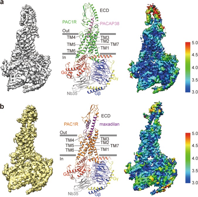
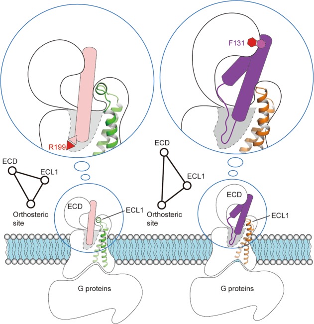
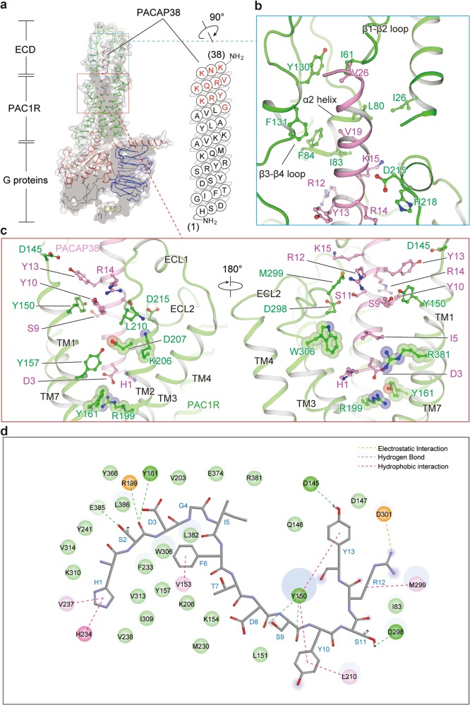
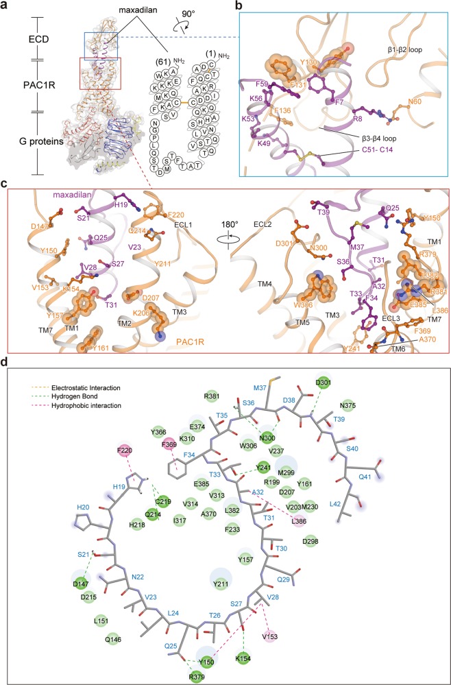
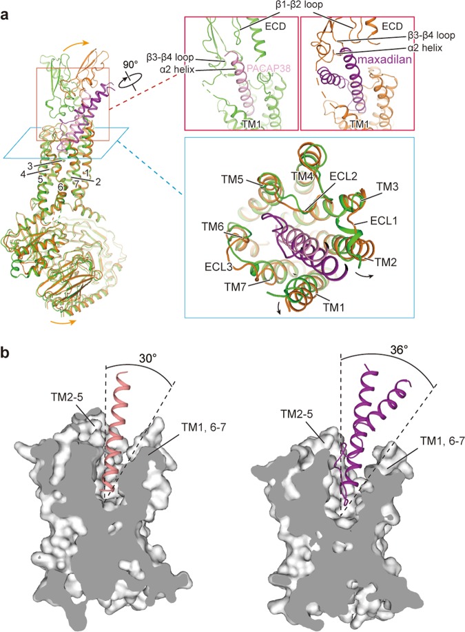
The pituitary adenylate cyclase-activating polypeptide type I receptor (PAC1R) belongs to the secretin receptor family and is widely distributed in the central neural system and peripheral organs. Abnormal activation of the receptor mediates trigeminovascular activation and sensitization, which is highly related to migraine, making PAC1R a potential therapeutic target. Elucidation of PAC1R activation mechanism would benefit discovery of therapeutic drugs for neuronal disorders. PAC1R activity is governed by pituitary adenylate cyclase-activating polypeptide (PACAP), known as a major vasodilator neuropeptide, and maxadilan, a native peptide from the sand fly, which is also capable of activating the receptor with similar potency. These peptide ligands have divergent sequences yet initiate convergent PAC1R activity. It is of interest to understand the mechanism of PAC1R ligand recognition and receptor activity regulation through structural biology. Here we report two near-atomic resolution cryo-EM structures of PAC1R activated by PACAP38 or maxadilan, providing structural insights into two distinct ligand binding modes. The structures illustrate flexibility of the extracellular domain (ECD) for ligands with distinct conformations, where ECD accommodates ligands in different orientations while extracellular loop 1 (ECL1) protrudes to further anchor the ligand bound in the orthosteric site. By structure-guided molecular modeling and mutagenesis, we tested residues in the ligand-binding pockets and identified clusters of residues that are critical for receptor activity. The structures reported here for the first time elucidate the mechanism of specificity and flexibility of ligand recognition and binding for PAC1R, and provide insights toward the design of therapeutic molecules targeting PAC1R.
垂体腺苷酸环化酶激活肽 I 型受体(PAC1R)属于分泌素受体家族,广泛分布于中枢神经系统和外周器官。该受体的异常激活介导三叉神经血管激活和敏化,这与偏头痛高度相关,使 PAC1R 成为潜在的治疗靶点。阐明 PAC1R 的激活机制将有助于发现治疗神经元疾病的治疗药物。PAC1R 的活性受垂体腺苷酸环化酶激活肽(PACAP)的调节,PACAP 是一种主要的血管舒张神经肽,以及沙蝇来源的内源性肽 maxadilan,它也具有激活受体的相似效力。这些肽配体具有不同的序列,但启动 PAC1R 活性的方式却趋同。通过结构生物学了解 PAC1R 配体识别和受体活性调节的机制是很有意义的。在这里,我们报告了两种接近原子分辨率的 PAC1R 冷冻电镜结构,分别被 PACAP38 或 maxadilan 激活,为两种不同的配体结合模式提供了结构见解。这些结构说明了配体具有不同构象时,细胞外结构域(ECD)的灵活性,其中 ECD 以不同的取向容纳配体,而细胞外环 1(ECL1)突出以进一步固定在正位结合点结合的配体。通过结构引导的分子建模和突变分析,我们测试了配体结合口袋中的残基,并确定了对受体活性至关重要的残基簇。这里报道的结构首次阐明了 PAC1R 识别和结合配体特异性和灵活性的机制,并为设计针对 PAC1R 的治疗分子提供了思路。