• 文献检索
  • 文档翻译
  • 深度研究
  • 学术资讯
  • Suppr Zotero 插件Zotero 插件
  • 邀请有礼
  • 套餐&价格
  • 历史记录
应用&插件
Suppr Zotero 插件Zotero 插件浏览器插件Mac 客户端Windows 客户端微信小程序
定价
高级版会员购买积分包购买API积分包
服务
文献检索文档翻译深度研究API 文档MCP 服务
关于我们
关于 Suppr公司介绍联系我们用户协议隐私条款
关注我们

Suppr 超能文献

核心技术专利:CN118964589B侵权必究
粤ICP备2023148730 号-1Suppr @ 2026

文献检索

告别复杂PubMed语法,用中文像聊天一样搜索,搜遍4000万医学文献。AI智能推荐,让科研检索更轻松。

立即免费搜索

文件翻译

保留排版,准确专业,支持PDF/Word/PPT等文件格式,支持 12+语言互译。

免费翻译文档

深度研究

AI帮你快速写综述,25分钟生成高质量综述,智能提取关键信息,辅助科研写作。

立即免费体验

根管系统及其分支的可变性和形态特征的微计算机断层评估。

Micro-computed tomographic assessment of the variability and morphological features of root canal system and their ramifications.

机构信息

Universidade de São Paulo, Faculdade de Odontologia de Ribeirão Preto, Departamento de Odontologia Restauradora, Ribeirão Preto, São Paulo, Brasil.

University of Leuven, Faculty of Medicine, Departament of Imaging and Pathology, OMFS IMPATH Research Group, Leuven, Belgium.

出版信息

J Appl Oral Sci. 2020 Feb 7;28:e20190393. doi: 10.1590/1678-7757-2019-0393. eCollection 2020.

DOI:10.1590/1678-7757-2019-0393
PMID:32049137
原文链接:https://pmc.ncbi.nlm.nih.gov/articles/PMC6999120/
Abstract

OBJECTIVES

This study assessed the incidence and variability features of root canals system (RCS) and their ramifications according to Pucci & Reig (PR) (1944) and the American Association of Endodontists (AAE) (2017) by micro-computed tomography (μCT).

METHODOLOGY

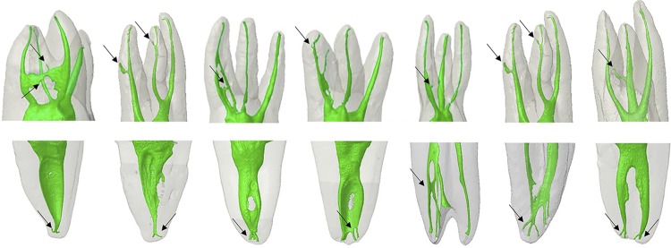
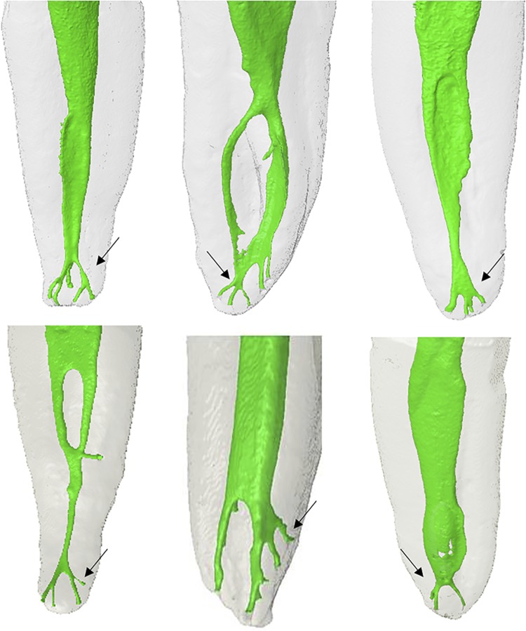
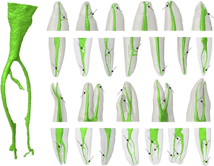
500 representative extracted human teeth of each tooth group (n=50) (maxillary/mandibular central and lateral incisors, canines, first and second premolars and molars) were scanned by μCT with a resolution of 26.70 μm. The reconstructed cross-sections images and the visualization of the continuous slices in the transversal axis were performed using DataViewer software. RCS were classified according to Pucci & Reig (main canal, collateral canal, lateral canal, secondary canal, accessory canal, intercanal, recurrent canal) and AAE (main canal, accessory canal, lateral canal). The apical deltas were assessed for both classifications. The prevalence of apical deltas was evaluated using the Chi-squared test (p<0.05).

RESULTS

According to PR, a higher incidence of lateral canals was observed in maxillary canines (10%), central incisors (8%) and first premolars (6%). Using AAE, the highest incidence of lateral canals was observed in the mandibular first premolars (85%), first and second molars (84%), lateral incisors (67%), canines (59%), and in maxillary first premolars (52%). Regarding accessory canals, the PR showed a frequency in 2% of the maxillary lateral incisors and maxillary and mandibular first premolars and 3% of mandibular first and second molars. On the other hand, the AAE showed the highest incidence of accessory canals in 86% of the maxillary first premolars, 71% in mandibular lateral incisors, 69% in mandibular first premolars, 65% in mandibular canines, and 56% in maxillary canines. The PR showed the lowest incidence of apical deltas for all dental groups when compared with AAE (p=0.004). Interestingly, distal canals in maxillary molars showed a significant discrepancy between classifications (p=0.027).

CONCLUSIONS

μCT enabled accurately describing the RC system and related ramifications, adding to the PR and AAE classifications, with some discrepancies reported for maxillary molars. Clinical Relevance This μCT study enabled a thorough description of the variability among root canals and their ramifications, including clinically relevant details on the presence and location of lateral canals and accessories in all human tooth groups, beyond the currently existing classification systems.

摘要

目的

本研究通过微计算机断层扫描(μCT)评估 Pucci & Reig(PR)(1944 年)和美国牙髓病学会(AAE)(2017 年)根管系统(RCS)及其分支的发生率和变异性特征。

方法

每组(上颌/下颌中切牙、侧切牙、尖牙、第一和第二前磨牙、磨牙)各随机抽取 50 颗有代表性的人离体牙(n=50),使用分辨率为 26.70 μm 的 μCT 进行扫描。使用 DataViewer 软件对重建的横截面图像和横轴上的连续切片进行可视化处理。根据 Pucci & Reig(主根管、侧副根管、侧根管、次根管、副根管、根管间、再通根管)和 AAE(主根管、副根管、侧根管)对 RCS 进行分类。评估两种分类的根尖差异。使用卡方检验(p<0.05)评估根尖差异的发生率。

结果

根据 PR,上颌尖牙(10%)、中切牙(8%)和第一前磨牙(6%)的侧根管发生率较高。根据 AAE,下颌第一前磨牙(85%)、第一和第二磨牙(84%)、侧切牙(67%)、尖牙(59%)和上颌第一前磨牙(52%)的侧根管发生率最高。关于副根管,PR 在 2%的上颌侧切牙和上颌及下颌第一前磨牙以及 3%的下颌第一和第二磨牙中显示出频率。另一方面,AAE 显示上颌第一前磨牙的副根管发生率最高(86%),下颌侧切牙(71%)、下颌第一前磨牙(69%)、下颌尖牙(65%)和上颌尖牙(56%)。与 AAE 相比,PR 显示所有牙组的根尖差异发生率最低(p=0.004)。有趣的是,上颌磨牙的远中根管在两种分类之间存在显著差异(p=0.027)。

结论

μCT 能够准确描述根管系统及其分支,与 PR 和 AAE 分类相比,增加了一些上颌磨牙的差异。

临床意义

本 μCT 研究对根管及其分支的变异性进行了全面描述,包括在所有人类牙组中有关侧根管和副根管的临床相关细节,包括其存在和位置,超出了目前现有的分类系统。

https://cdn.ncbi.nlm.nih.gov/pmc/blobs/e6ff/6999120/7d6e0269ed37/1678-7757-jaos-28-e20190393-gf03.jpg
https://cdn.ncbi.nlm.nih.gov/pmc/blobs/e6ff/6999120/6b33ed0a510a/1678-7757-jaos-28-e20190393-gf01.jpg
https://cdn.ncbi.nlm.nih.gov/pmc/blobs/e6ff/6999120/212583520b5f/1678-7757-jaos-28-e20190393-gf02.jpg
https://cdn.ncbi.nlm.nih.gov/pmc/blobs/e6ff/6999120/7d6e0269ed37/1678-7757-jaos-28-e20190393-gf03.jpg

相似文献

1
Micro-computed tomographic assessment of the variability and morphological features of root canal system and their ramifications.根管系统及其分支的可变性和形态特征的微计算机断层评估。
J Appl Oral Sci. 2020 Feb 7;28:e20190393. doi: 10.1590/1678-7757-2019-0393. eCollection 2020.
2
A stereomicroscopy study of root apices of human maxillary central incisors and mandibular second premolars in an Iranian population.伊朗人群中人类上颌中切牙和下颌第二前磨牙根尖的体视显微镜研究。
J Oral Sci. 2009 Sep;51(3):411-5. doi: 10.2334/josnusd.51.411.
3
Micro-Computed Tomography Analysis of the Root Canal Morphology of Palatal Roots of Maxillary First Molars.上颌第一磨牙腭根根管形态的显微计算机断层扫描分析
J Endod. 2016 Feb;42(2):280-3. doi: 10.1016/j.joen.2015.10.016. Epub 2015 Nov 26.
4
Micro-computed tomographic analysis of the root canal morphology of the distal root of mandibular first molar.下颌第一磨牙远中根根管形态的显微计算机断层扫描分析
J Endod. 2015 Feb;41(2):231-6. doi: 10.1016/j.joen.2014.09.024. Epub 2014 Nov 12.
5
Root canal morphology of permanent teeth in a Malaysian subpopulation using cone-beam computed tomography.马来西亚人群恒牙根尖形态的锥形束 CT 研究。
BMC Oral Health. 2019 Jan 14;19(1):14. doi: 10.1186/s12903-019-0710-z.
6
Micro-computed tomographic analysis of the root canal morphology of mandibular incisors.下颌切牙根管形态的显微计算机断层扫描分析
J Endod. 2014 May;40(5):710-6. doi: 10.1016/j.joen.2013.09.003. Epub 2013 Oct 16.
7
A micro-computed tomographic analysis of the root canal systems in the permanent mandibular incisors in a Chinese population.中国人恒牙下颌切牙根管系统的微计算机断层分析。
BMC Oral Health. 2023 Mar 8;23(1):129. doi: 10.1186/s12903-023-02830-5.
8
Impacts of conservative endodontic cavity on root canal instrumentation efficacy and resistance to fracture assessed in incisors, premolars, and molars.保守性牙髓治疗窝洞对切牙、前磨牙和磨牙根管预备效果及抗折性的影响评估
J Endod. 2014 Aug;40(8):1160-6. doi: 10.1016/j.joen.2013.12.012. Epub 2014 Jan 28.
9
Morphologic micro-computed tomography analysis of mandibular premolars with three root canals.具有三根根管的下颌前磨牙的形态学显微计算机断层扫描分析
J Endod. 2013 Sep;39(9):1130-5. doi: 10.1016/j.joen.2013.02.007. Epub 2013 Apr 10.
10
Incidence of accessory canals in Japanese anterior maxillary teeth following root canal filling ex vivo.日本上颌前牙根管充填后副根管的发生率。
Int Endod J. 2010 May;43(5):370-6. doi: 10.1111/j.1365-2591.2010.01688.x.

引用本文的文献

1
Aloe Vera as an Adjunct in Endodontic Irrigation: Impact on Dentin Bond Strength and Cytotoxicity.芦荟作为根管冲洗的辅助剂:对牙本质粘结强度和细胞毒性的影响。
Materials (Basel). 2025 Jun 18;18(12):2874. doi: 10.3390/ma18122874.
2
A unique AI-based tool for automated segmentation of pulp cavity structures in maxillary premolars on CBCT.一种基于人工智能的独特工具,用于在上颌前磨牙的CBCT上自动分割牙髓腔结构。
Sci Rep. 2025 Feb 14;15(1):5509. doi: 10.1038/s41598-025-86203-8.
3
Description of the root anatomy of the primary molars using high resolution computed microtomography (Micro-CT). An analysis of three-dimensional root canal system.

本文引用的文献

1
Micro-computed Tomographic Evaluation of the Prevalence, Distribution, and Morphologic Features of Accessory Canals in Chinese Permanent Teeth.中国人恒牙中额外管的发生率、分布及形态特征的 micro-computed tomographic 评估。
J Endod. 2019 Aug;45(8):994-999. doi: 10.1016/j.joen.2019.04.001. Epub 2019 Jun 13.
2
Apical root canal anatomy in the mesiobuccal root of maxillary first molars: influence of root apical shape and prevalence of apical foramina - a micro-CT study.上颌第一磨牙近中颊根的根尖根管解剖结构:根尖形态和根尖孔发生率的影响——一项显微 CT 研究。
Int Endod J. 2019 Aug;52(8):1218-1227. doi: 10.1111/iej.13109. Epub 2019 Mar 28.
3
使用高分辨率计算机显微断层扫描(Micro-CT)对乳磨牙牙根解剖结构进行描述。三维根管系统分析。
Front Dent Med. 2025 Jan 6;5:1522414. doi: 10.3389/fdmed.2024.1522414. eCollection 2024.
4
A novel artificial intelligence-powered tool for automated root canal segmentation in single-rooted teeth on cone-beam computed tomography.一种新型的人工智能驱动工具,用于在锥形束计算机断层扫描上对单根牙进行自动根管分割。
Int Endod J. 2025 Apr;58(4):658-671. doi: 10.1111/iej.14200. Epub 2025 Jan 28.
5
Comparative Evaluation of Different Obturation Techniques for Root Canal Filling of Permanent Teeth: An In-Vitro Study.恒牙根管充填不同充填技术的比较评价:一项体外研究
Cureus. 2024 Sep 4;16(9):e68623. doi: 10.7759/cureus.68623. eCollection 2024 Sep.
6
Negotiating and Treating Lateral Canals: Report of 7 Cases.侧隐窝狭窄的诊治:附 7 例报告
Eur Endod J. 2024 Aug 22;9(3):287-294. doi: 10.14744/eej.2023.43265.
7
Root canal morphology of permanent mandibular anterior teeth in a Pakistani population: A cone beam computed tomography assessment.巴基斯坦人群中恒牙下颌前牙的根管形态:锥形束计算机断层扫描评估
Pak J Med Sci. 2024 Aug;40(7):1529-1532. doi: 10.12669/pjms.40.7.8744.
8
Micro-CT and histological examination of accessory canals in 34 equine cheek teeth.34颗马颊齿副根管的显微CT及组织学检查
Front Vet Sci. 2024 Apr 10;11:1396871. doi: 10.3389/fvets.2024.1396871. eCollection 2024.
9
Diagnostic value of cone beam computed tomography for root canal morphology assessment - a micro-CT based comparison.锥形束计算机断层扫描在根管形态评估中的诊断价值——基于微 CT 的比较。
Clin Oral Investig. 2024 Mar 8;28(3):201. doi: 10.1007/s00784-024-05580-y.
10
Assessment of the Effect of Different Irrigation Protocols on the Penetration of Irrigation Solution into Simulated Lateral Canals (In Vitro Study).不同冲洗方案对模拟侧支根管内冲洗液渗透效果的评估(体外研究)。
Eur Endod J. 2024 Mar 8;9(2):146 - 153. doi: 10.14744/eej.2023.29494. Epub 2024 Jan 12.
Evaluation of the Root Canal Anatomy of Maxillary and Mandibular Premolars in a Selected German Population Using Cone-beam Computed Tomographic Data.
利用锥形束 CT 数据评估选定德国人群上颌和下颌前磨牙的根管解剖结构。
J Endod. 2017 Sep;43(9):1448-1452. doi: 10.1016/j.joen.2017.03.044. Epub 2017 Jul 22.
4
Dentinal Microcrack Development after Canal Preparation: A Longitudinal in Situ Micro-computed Tomography Study Using a Cadaver Model.根管预备后牙本质微裂纹的发展:使用尸体模型的纵向原位微计算机断层扫描研究。
J Endod. 2017 Sep;43(9):1553-1558. doi: 10.1016/j.joen.2017.04.027. Epub 2017 Jul 20.
5
A new system for classifying accessory canal morphology.一种新的副根管形态分类系统。
Int Endod J. 2018 Feb;51(2):164-176. doi: 10.1111/iej.12800. Epub 2017 Jul 31.
6
The Use of a Novel Approach for the Instrumentation of a Cone-beam Computed Tomography-discernible Lateral Canal in an Unusual Maxillary Incisor: Case Report.一种用于上颌中切牙异常情况下锥束计算机断层扫描可识别的侧根管器械操作的新方法:病例报告
J Endod. 2017 Jun;43(6):1023-1027. doi: 10.1016/j.joen.2017.01.032. Epub 2017 Apr 14.
7
Morphological evaluation of maxillary second molars with fused roots: a micro-CT study.上颌第二磨牙融合根的形态学评估:一项微 CT 研究。
Int Endod J. 2017 Dec;50(12):1192-1200. doi: 10.1111/iej.12752. Epub 2017 Mar 10.
8
Root canal morphology and configuration of 123 maxillary second molars by means of micro-CT.上颌第二磨牙 123 根管形态和解剖结构的 micro-CT 研究。
Int J Oral Sci. 2017 Mar;9(1):33-37. doi: 10.1038/ijos.2016.53. Epub 2017 Jan 20.
9
Unusual Deviation of the Main Foramen from the Root Apex.主孔与根尖的异常偏离。
Braz Dent J. 2016 Sep-Oct;27(5):589-591. doi: 10.1590/0103-6440201601106.
10
Micro-CT evaluation of apical delta morphologies in human teeth.人牙牙尖三角形态的 micro-CT 评估
Sci Rep. 2016 Nov 7;6:36501. doi: 10.1038/srep36501.