Division of Advanced Surgical Science and Technology, Tohoku University Graduate School of Medicine, Sendai, Miyagi, Japan.
Division of Gastroenterologic and Hepatobiliarypancreatic Surgery, Tohoku Medical and Pharmaceutical University Hospital, Sendai, Miyagi, Japan.
Sci Rep. 2020 Feb 12;10(1):2405. doi: 10.1038/s41598-020-58369-w.
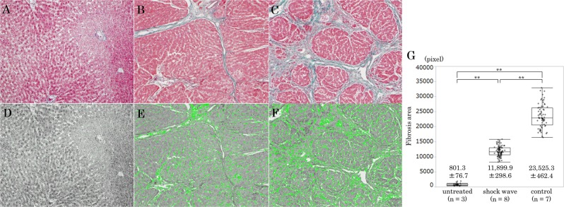
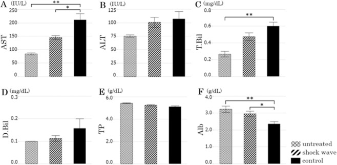
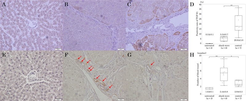
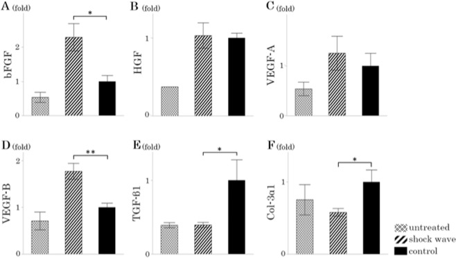
Low-energy extracorporeal shock waves (LESW) have been studied as a new treatment for angina pectoris and several ischemic diseases because of its effect on angiogenesis and inhibition of fibrosis of the heart. The effect of LESW on fibrosis in liver cirrhosis has not been studied. The aim of this study was to verify the amelioration of liver fibrosis by LESW and elucidate its mechanisms in a rat model of drug-induced liver cirrhosis. Male Wistar rats aged 7 weeks were injected with carbon tetrachloride intraperitoneally twice a week for 12 weeks. Eight rats underwent LESW therapy (0.25 mJ/mm, 4 Hz, 1000 shots) under general anesthesia (shock wave group). Seven rats only underwent general anesthesia (control group). Quantitative analysis showed that the area of fibrosis in the shock wave group was significantly reduced compared with the control group (11,899.9 vs. 23,525.3 pixels per field, p < 0.001). In the shock wave group, the mRNA expression of transforming growth factor (TGF)-β1 was significantly suppressed (0.40-fold, p = 0.018) and vascular endothelial growth factor-B was significantly increased (1.77-fold, p = 0.006) compared with the control group. Serum albumin was significantly higher in the shock wave group than in the control group (3.0 vs. 2.4 g/dl, p = 0.025). Aspartate aminotransferase/alanine aminotransferase ratio decreased by LESW compared with the control group (1.49 vs. 2.04, p = 0.013). These results suggest that LESW therapy ameliorates liver fibrosis by reducing the expression of TGF-β1 and increasing the expression of angiogenic factors, and improves hepatic function.
低能量体外冲击波(LESW)已被研究作为一种新的治疗心绞痛和几种缺血性疾病的方法,因为它对血管生成和心脏纤维化的抑制作用。LESW 对肝硬化纤维化的影响尚未得到研究。本研究旨在验证 LESW 对肝硬化纤维化的改善,并在药物诱导的肝硬化大鼠模型中阐明其机制。7 周龄雄性 Wistar 大鼠每周两次经腹腔注射四氯化碳 12 周。8 只大鼠在全身麻醉下接受 LESW 治疗(0.25mJ/mm,4Hz,1000 次)(冲击波组)。7 只大鼠仅接受全身麻醉(对照组)。定量分析显示,冲击波组的纤维化面积明显小于对照组(11899.9 与 23525.3 像素/视野,p<0.001)。在冲击波组,转化生长因子(TGF)-β1 的 mRNA 表达明显受到抑制(0.40 倍,p=0.018),血管内皮生长因子-B 明显增加(1.77 倍,p=0.006)。与对照组相比,冲击波组血清白蛋白明显升高(3.0 与 2.4g/dl,p=0.025)。与对照组相比,LESW 降低了天冬氨酸氨基转移酶/丙氨酸氨基转移酶比值(1.49 与 2.04,p=0.013)。这些结果表明,LESW 治疗通过降低 TGF-β1 的表达和增加血管生成因子的表达来改善肝纤维化,并改善肝功能。