Lung Cellular and Molecular Biology Laboratory, Institute of Pulmonary Medicine, Hadassah Medical Research Center, PO Box 12000, Kiryat Hadassah, Jerusalem 91120, Israel.
Department of Pathology and Laboratory Medicine, 670 Albany St, 4th Floor, Boston University School of Medicine, Boston, MA 02118, USA.
Cells. 2020 Feb 11;9(2):411. doi: 10.3390/cells9020411.
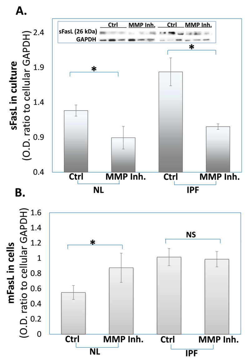
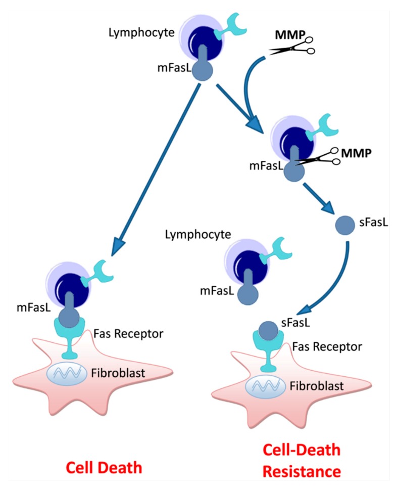
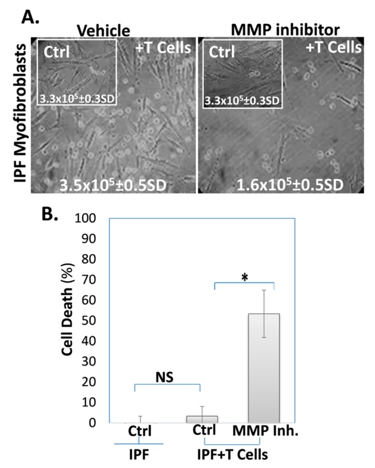
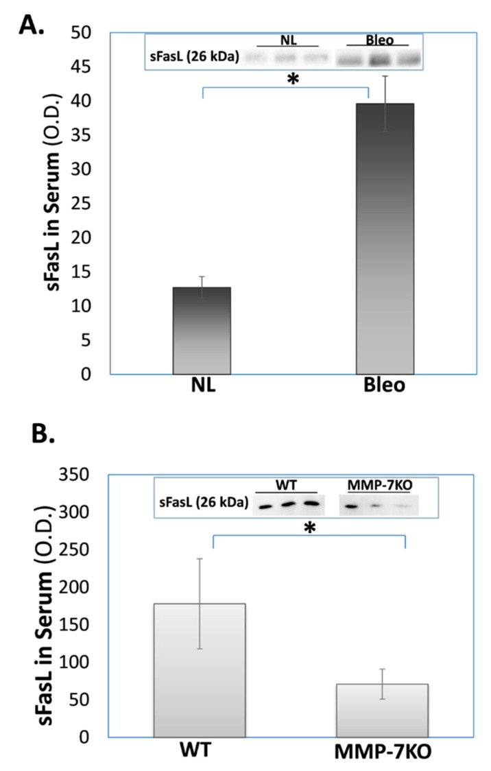
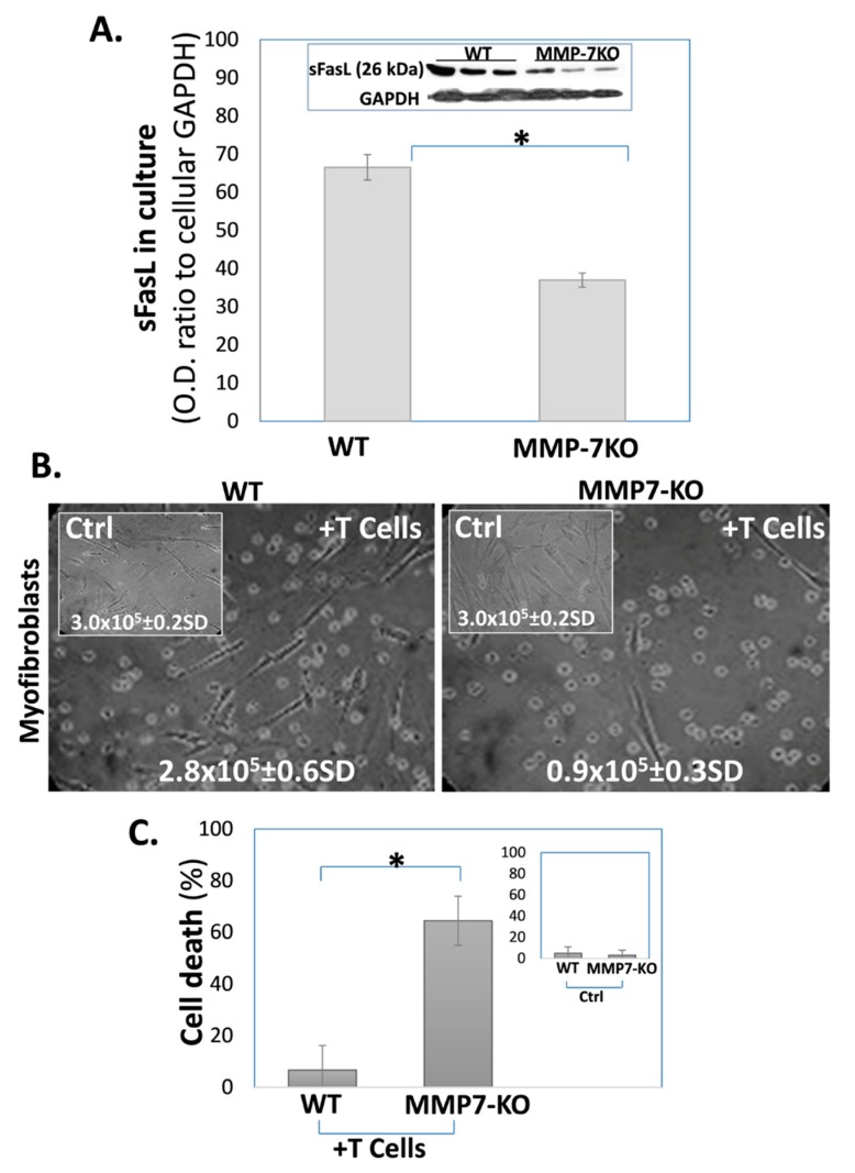
A prominent feature of obstructed tissue regeneration following injury in general, and fibrotic lung tissue in particular, is fibroblast proliferation and accumulation. The Fas/FasL apoptotic pathway has been shown to be involved in human idiopathic pulmonary fibrosis (IPF) and bleomycin-induced lung fibrosis in rodents. We previously showed that in normal injury repair, myofibroblasts' accumulation is followed by their decline by FasL T cell-induced cell death. In pathological lung fibrosis, myofibroblasts resist cell death and accumulate. Like other members of the tumor necrosis factor (TNF) family, membrane-bound FasL can be cleaved from the cell surface to generate a soluble form (sFasL). Metalloproteinases (MMPs) are known to convert the membrane-bound form of FasL to sFasL. MMP-7 knockout (KO) mice were shown to be protected from bleomycin (BLM)-induced lung fibrosis. In this study, we detected increased levels of sFasL in their blood serum, as in the lungs of patients with IPF, and IPF-lung myofibroblast culture medium. In this study, using an MMP-inhibitor, we showed that sFasL is decreased in cultures of IPF-lung myofibroblasts and BLM-treated lung myofibroblasts, and in the blood serum of MMP-7KO mice. Moreover, resistant fibrotic-lung myofibroblasts, from the lungs of humans with IPF and of BLM-treated mice, became susceptible to T-cell induced cell death in a co-culture following MMP-inhibition- vs. control-treatment or BLM-treated MMP-7KO vs. wild-type mice, respectively. sFasL may be an unrecognized mechanism for MMP-7-mediated decreased tissue regeneration following injury and the evolution of lung fibrosis.
一般来说,组织损伤后再生受阻的一个显著特征,特别是纤维化肺组织,是成纤维细胞的增殖和积累。Fas/FasL 凋亡途径已被证明参与人类特发性肺纤维化 (IPF) 和博莱霉素诱导的啮齿动物肺纤维化。我们之前表明,在正常的损伤修复中,肌成纤维细胞的积累随后被 FasL T 细胞诱导的细胞死亡所减少。在病理性肺纤维化中,肌成纤维细胞抵抗细胞死亡并积累。像肿瘤坏死因子 (TNF) 家族的其他成员一样,膜结合 FasL 可以从细胞表面被切割产生可溶性形式 (sFasL)。已知金属蛋白酶 (MMP) 将 FasL 的膜结合形式转化为 sFasL。MMP-7 敲除 (KO) 小鼠被证明可以免受博莱霉素 (BLM) 诱导的肺纤维化的影响。在这项研究中,我们在血清中检测到了与 IPF 患者和 IPF 肺成纤维细胞培养物中相同的 sFasL 水平升高。在这项研究中,我们使用 MMP 抑制剂表明,sFasL 在 IPF 肺成纤维细胞和 BLM 处理的肺成纤维细胞的培养物以及 MMP-7KO 小鼠的血清中减少。此外,来自 IPF 患者和 BLM 处理的小鼠的耐药性纤维化肺成纤维细胞,在 MMP 抑制与对照处理或 BLM 处理的 MMP-7KO 与野生型小鼠的共培养后,对 T 细胞诱导的细胞死亡变得敏感。sFasL 可能是 MMP-7 介导的损伤后组织再生减少和肺纤维化进展的未被认识的机制。