• 文献检索
  • 文档翻译
  • 深度研究
  • 学术资讯
  • Suppr Zotero 插件Zotero 插件
  • 邀请有礼
  • 套餐&价格
  • 历史记录
应用&插件
Suppr Zotero 插件Zotero 插件浏览器插件Mac 客户端Windows 客户端微信小程序
定价
高级版会员购买积分包购买API积分包
服务
文献检索文档翻译深度研究API 文档MCP 服务
关于我们
关于 Suppr公司介绍联系我们用户协议隐私条款
关注我们

Suppr 超能文献

核心技术专利:CN118964589B侵权必究
粤ICP备2023148730 号-1Suppr @ 2026

文献检索

告别复杂PubMed语法,用中文像聊天一样搜索,搜遍4000万医学文献。AI智能推荐,让科研检索更轻松。

立即免费搜索

文件翻译

保留排版,准确专业,支持PDF/Word/PPT等文件格式,支持 12+语言互译。

免费翻译文档

深度研究

AI帮你快速写综述,25分钟生成高质量综述,智能提取关键信息,辅助科研写作。

立即免费体验

免疫磁分离-电化学免疫传感法分离乳腺癌来源的纳米囊泡中的基质效应:比较研究。

Matrix Effect in the Isolation of Breast Cancer-Derived Nanovesicles by Immunomagnetic Separation and Electrochemical Immunosensing-A Comparative Study.

机构信息

Grup de Sensors i Biosensors, Departament de Química, Universitat Autònoma de Barcelona, 08193 Bellaterra, Spain.

Laboratori d'Immunologia Cel·lular, Institut de Biotecnologia i Biomedicina, Universitat Autònoma de Barcelona, 08193 Bellaterra, Spain.

出版信息

Sensors (Basel). 2020 Feb 11;20(4):965. doi: 10.3390/s20040965.

DOI:10.3390/s20040965
PMID:32054015
原文链接:https://pmc.ncbi.nlm.nih.gov/articles/PMC7071381/
Abstract

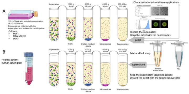
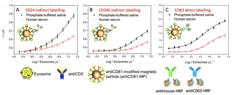
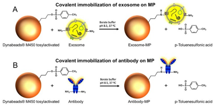
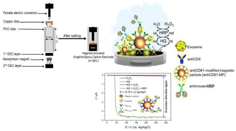
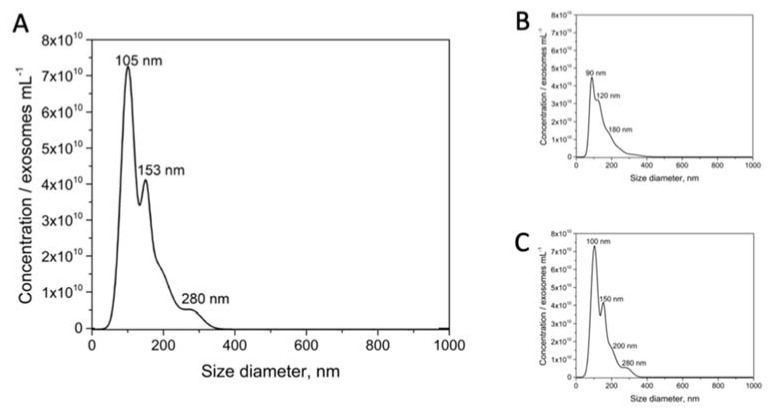
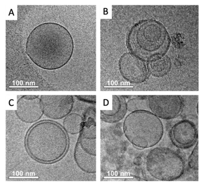
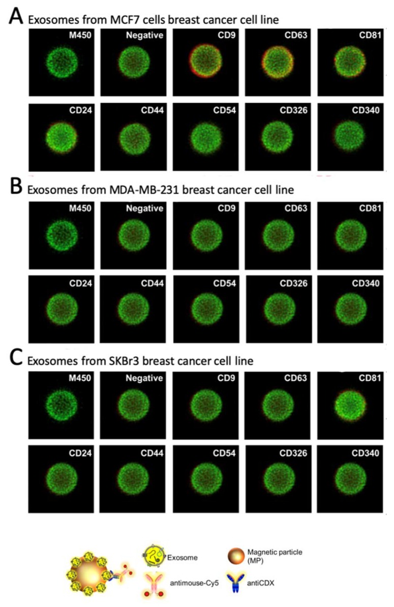
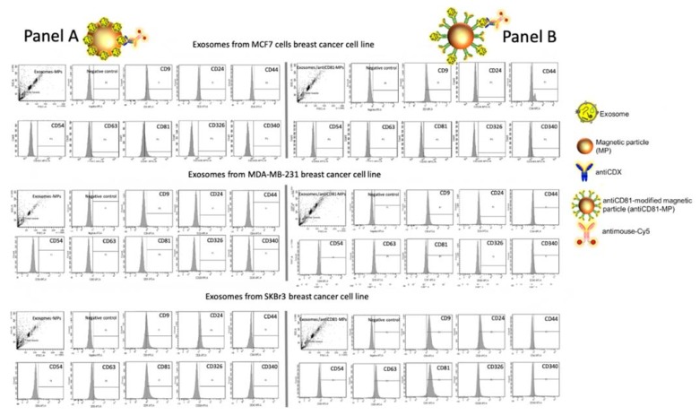
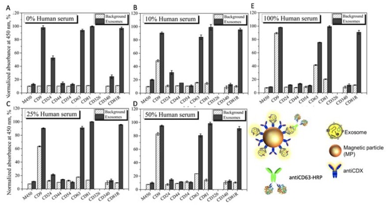
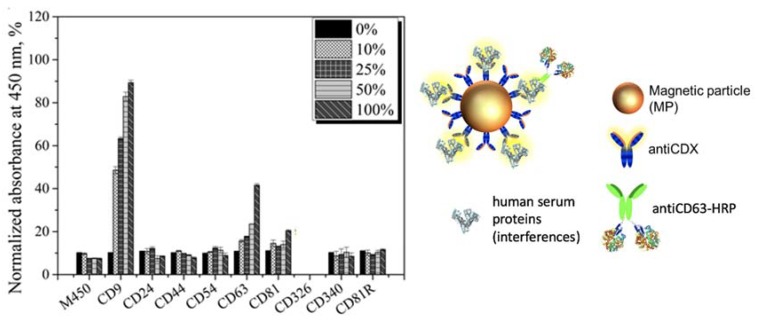
Exosomes are cell-derived nanovesicles released into biological fluids, which are involved in cell-to-cell communication. The analysis of the content and the surface of the exosomes allow conclusions about the cells they are originating from and the underlying condition, pathology or disease. Therefore, the exosomes are currently considered good candidates as biomarkers to improve the current methods for clinical diagnosis, including cancer. However, due to their low concentration, conventional procedures for exosome detection including biosensing usually require relatively large sample volumes and involve preliminary purification and preconcentration steps by ultracentrifugation. In this paper, the immunomagnetic separation is presented as an alternative method for the specific isolation of exosomes in serum. To achieve that, a rational study of the surface proteins in exosomes, which can be recognized by magnetic particles, is presented. The characterization was performed in exosomes obtained from cell culture supernatants of MCF7, MDA-MB-231 and SKBR3 breast cancer cell lines, including TEM and nanoparticle tracking analysis (NTA). For the specific characterization by flow cytometry and confocal microscopy, different commercial antibodies against selected receptors were used, including the general tetraspanins CD9, CD63 and CD81, and cancer-related receptors (CD24, CD44, CD54, CD326 and CD340). The effect of the serum matrix on the immunomagnetic separation was then carefully evaluated by spiking the exosomes in depleted human serum. Based on this study, the exosomes were preconcentrated by immunomagnetic separation on antiCD81-modified magnetic particles in order to achieve further magnetic actuation on the surface of the electrode for the electrochemical readout. The performance of this approach is discussed and compared with classical characterization methods.

摘要

外泌体是一种细胞衍生的纳米囊泡,释放到生物体液中,参与细胞间通讯。对外泌体的内容物和表面进行分析,可以得出它们所源自的细胞以及潜在的情况、病理学或疾病的结论。因此,外泌体目前被认为是改善当前临床诊断方法(包括癌症)的良好生物标志物候选物。然而,由于其浓度较低,包括用于癌症检测的生物传感器在内的常规外泌体检测方法通常需要相对较大的样本量,并涉及通过超速离心进行初步的纯化和预浓缩步骤。在本文中,免疫磁分离被提出作为一种替代方法,用于在血清中特异性分离外泌体。为了实现这一目标,本文对外泌体表面蛋白进行了合理的研究,这些蛋白可以被磁性颗粒识别。通过 MCF7、MDA-MB-231 和 SKBR3 乳腺癌细胞系的细胞培养上清液中获得的外泌体进行了 TEM 和纳米颗粒跟踪分析 (NTA) 等表征。为了通过流式细胞术和共聚焦显微镜进行特异性表征,使用了不同的针对选定受体的商业抗体,包括通用四跨膜蛋白 CD9、CD63 和 CD81,以及与癌症相关的受体(CD24、CD44、CD54、CD326 和 CD340)。然后,通过在耗尽的人血清中掺入外泌体,仔细评估了血清基质对外泌体免疫磁分离的影响。在此基础上,通过免疫磁分离在外泌体上进行预浓缩,以实现进一步的磁性作用,用于在外泌体表面的电极上进行电化学读出。讨论了这种方法的性能,并与经典的表征方法进行了比较。

https://cdn.ncbi.nlm.nih.gov/pmc/blobs/c7c7/7071381/d0147cc22974/sensors-20-00965-g010.jpg
https://cdn.ncbi.nlm.nih.gov/pmc/blobs/c7c7/7071381/aca6e9940e81/sensors-20-00965-g001.jpg
https://cdn.ncbi.nlm.nih.gov/pmc/blobs/c7c7/7071381/b25ce18da8af/sensors-20-00965-g002.jpg
https://cdn.ncbi.nlm.nih.gov/pmc/blobs/c7c7/7071381/387523368867/sensors-20-00965-g003.jpg
https://cdn.ncbi.nlm.nih.gov/pmc/blobs/c7c7/7071381/8ebfab1c4f36/sensors-20-00965-g004.jpg
https://cdn.ncbi.nlm.nih.gov/pmc/blobs/c7c7/7071381/78462ce8ba45/sensors-20-00965-g005.jpg
https://cdn.ncbi.nlm.nih.gov/pmc/blobs/c7c7/7071381/a9129b10adea/sensors-20-00965-g006.jpg
https://cdn.ncbi.nlm.nih.gov/pmc/blobs/c7c7/7071381/fd872963577c/sensors-20-00965-g007.jpg
https://cdn.ncbi.nlm.nih.gov/pmc/blobs/c7c7/7071381/5b7240c25f5e/sensors-20-00965-g008.jpg
https://cdn.ncbi.nlm.nih.gov/pmc/blobs/c7c7/7071381/75c8d7e0a3b1/sensors-20-00965-g009.jpg
https://cdn.ncbi.nlm.nih.gov/pmc/blobs/c7c7/7071381/d0147cc22974/sensors-20-00965-g010.jpg

相似文献

1
Matrix Effect in the Isolation of Breast Cancer-Derived Nanovesicles by Immunomagnetic Separation and Electrochemical Immunosensing-A Comparative Study.免疫磁分离-电化学免疫传感法分离乳腺癌来源的纳米囊泡中的基质效应:比较研究。
Sensors (Basel). 2020 Feb 11;20(4):965. doi: 10.3390/s20040965.
2
Electrochemical immunosensing of nanovesicles as biomarkers for breast cancer.电化学免疫传感纳米囊泡作为乳腺癌生物标志物。
Biosens Bioelectron. 2020 Feb 15;150:111882. doi: 10.1016/j.bios.2019.111882. Epub 2019 Nov 13.
3
Multiplex detection and characterization of breast cancer exosomes by magneto-actuated immunoassay.磁驱动免疫分析多指标检测和鉴定乳腺癌外泌体。
Talanta. 2020 May 1;211:120657. doi: 10.1016/j.talanta.2019.120657. Epub 2019 Dec 24.
4
Magnetic Separation of Cell-Secreted Vesicles with Tailored Magnetic Particles and Downstream Applications.定制磁性颗粒的细胞分泌囊泡的磁分离及其下游应用。
Methods Mol Biol. 2023;2668:257-276. doi: 10.1007/978-1-0716-3203-1_18.
5
Expression of B-cell surface antigens in subpopulations of exosomes released from B-cell lymphoma cells.B细胞淋巴瘤细胞释放的外泌体亚群中B细胞表面抗原的表达
Clin Ther. 2014 Jun 1;36(6):847-862.e1. doi: 10.1016/j.clinthera.2014.05.010.
6
Magnetic bead-based isolation of exosomes.基于磁珠的外泌体分离
Methods Mol Biol. 2015;1218:465-81. doi: 10.1007/978-1-4939-1538-5_27.
7
Exosomes from human placenta purified by affinity chromatography on sepharose bearing immobilized antibodies against CD81 tetraspanin contain many peptides and small proteins.用固定在 sepharose 上的针对 CD81 四跨膜蛋白的抗体亲和层析法从人胎盘中纯化的外泌体含有许多肽和小蛋白。
IUBMB Life. 2018 Nov;70(11):1144-1155. doi: 10.1002/iub.1928. Epub 2018 Oct 2.
8
Electrochemical Genosensing of Overexpressed GAPDH Transcripts in Breast Cancer Exosomes.电化学基因传感在乳腺癌外泌体中过表达的 GAPDH 转录本。
Anal Chem. 2023 Jan 31;95(4):2487-2495. doi: 10.1021/acs.analchem.2c04773. Epub 2023 Jan 22.
9
Immunomagnetic separation of Salmonella with tailored magnetic micro and nanocarriers. A comparative study.用定制的磁性微纳载体免疫磁分离沙门氏菌。比较研究。
Talanta. 2015 Oct 1;143:198-204. doi: 10.1016/j.talanta.2015.05.035. Epub 2015 May 23.
10
The activity of alkaline phosphatase in breast cancer exosomes simplifies the biosensing design.乳腺癌外泌体中碱性磷酸酶的活性简化了生物传感设计。
Biosens Bioelectron. 2022 Feb 15;198:113826. doi: 10.1016/j.bios.2021.113826. Epub 2021 Nov 23.

引用本文的文献

1
Exosome-Based Therapeutics: A Natural Solution to Overcoming the Blood-Brain Barrier in Neurodegenerative Diseases.基于外泌体的疗法:克服神经退行性疾病中血脑屏障的天然解决方案。
MedComm (2020). 2025 Sep 12;6(9):e70386. doi: 10.1002/mco2.70386. eCollection 2025 Sep.
2
Mesenchymal stem cell-derived extracellular vesicles: current advances in preparation and therapeutic applications for neurological disorders.间充质干细胞衍生的细胞外囊泡:神经系统疾病制备与治疗应用的当前进展
Front Cell Dev Biol. 2025 Aug 18;13:1626996. doi: 10.3389/fcell.2025.1626996. eCollection 2025.
3
Role of exosomes in castration-resistant prostate cancer.

本文引用的文献

1
Multiplex detection and characterization of breast cancer exosomes by magneto-actuated immunoassay.磁驱动免疫分析多指标检测和鉴定乳腺癌外泌体。
Talanta. 2020 May 1;211:120657. doi: 10.1016/j.talanta.2019.120657. Epub 2019 Dec 24.
2
Comparative analysis of exosome isolation methods using culture supernatant for optimum yield, purity and downstream applications.比较不同方法提取培养上清液中外泌体的产量、纯度和下游应用效果。
Sci Rep. 2019 Mar 29;9(1):5335. doi: 10.1038/s41598-019-41800-2.
3
EpCAM and EpCAM circulating tumor cells in metastatic prostate and breast cancer patients.
外泌体在去势抵抗性前列腺癌中的作用。
Front Oncol. 2025 May 14;15:1498733. doi: 10.3389/fonc.2025.1498733. eCollection 2025.
4
Mesenchymal Stem Cell-Derived Exosomes Hold Promise in the Treatment of Diabetic Foot Ulcers.间充质干细胞衍生的外泌体在糖尿病足溃疡治疗中具有前景。
Int J Nanomedicine. 2025 May 6;20:5837-5857. doi: 10.2147/IJN.S516533. eCollection 2025.
5
Precision Fabrication and Optimization of Nanostructures for Exosome Detection via Surface-Enhanced Raman Spectroscopy.通过表面增强拉曼光谱法精确制造和优化用于外泌体检测的纳米结构
Nanomaterials (Basel). 2025 Feb 10;15(4):266. doi: 10.3390/nano15040266.
6
Embryotrophic effect of exogenous protein contained adipose-derived stem cell extracellular vesicles.脂肪来源干细胞外泌体中所含外源蛋白的胚胎营养作用。
J Anim Sci Biotechnol. 2024 Nov 3;15(1):145. doi: 10.1186/s40104-024-01106-4.
7
Circulating Tumor Cells in Cancer Diagnosis, Therapy, and Theranostics Applications: An Overview of Emerging Materials and Technologies.循环肿瘤细胞在癌症诊断、治疗及诊疗一体化应用中的研究进展:新型材料与技术综述
Curr Pharm Des. 2025;31(9):674-690. doi: 10.2174/0113816128328459241009191933.
8
Isolation and usage of exosomes in central nervous system diseases.中枢神经系统疾病中外泌体的分离与应用。
CNS Neurosci Ther. 2024 Mar;30(3):e14677. doi: 10.1111/cns.14677.
9
Circulating tumor biomarkers in early-stage breast cancer: characteristics, detection, and clinical developments.早期乳腺癌中的循环肿瘤生物标志物:特征、检测及临床进展
Front Oncol. 2023 Oct 24;13:1288077. doi: 10.3389/fonc.2023.1288077. eCollection 2023.
10
Extracellular vesicles, the emerging mirrors of brain physiopathology.细胞外囊泡,大脑病理生理学的新兴镜像。
Int J Biol Sci. 2023 Jan 1;19(3):721-743. doi: 10.7150/ijbs.79063. eCollection 2023.
转移性前列腺癌和乳腺癌患者中的上皮细胞黏附分子(EpCAM)及循环肿瘤细胞(EpCAM CTCs)
Oncotarget. 2018 Nov 2;9(86):35705-35716. doi: 10.18632/oncotarget.26298.
4
Epithelial cell adhesion molecule (EpCAM) is involved in prostate cancer chemotherapy/radiotherapy response in vivo.上皮细胞黏附分子(EpCAM)参与体内前列腺癌的化疗/放疗反应。
BMC Cancer. 2018 Nov 12;18(1):1092. doi: 10.1186/s12885-018-5010-5.
5
Clinico-pathological significance of exosome marker CD63 expression on cancer cells and stromal cells in gastric cancer.胃癌中癌细胞和基质细胞来源外泌体标志物 CD63 表达的临床病理意义。
PLoS One. 2018 Sep 17;13(9):e0202956. doi: 10.1371/journal.pone.0202956. eCollection 2018.
6
Global cancer statistics 2018: GLOBOCAN estimates of incidence and mortality worldwide for 36 cancers in 185 countries.全球癌症统计数据 2018:GLOBOCAN 对全球 185 个国家/地区 36 种癌症的发病率和死亡率的估计。
CA Cancer J Clin. 2018 Nov;68(6):394-424. doi: 10.3322/caac.21492. Epub 2018 Sep 12.
7
Increased Expression of CD81 in Breast Cancer Tissue is Associated with Reduced Patient Prognosis and Increased Cell Migration and Proliferation in MDA-MB-231 and MDA-MB-435S Human Breast Cancer Cell Lines In Vitro.乳腺癌组织中 CD81 的表达增加与患者预后降低以及 MDA-MB-231 和 MDA-MB-435S 人乳腺癌细胞系体外迁移和增殖增加有关。
Med Sci Monit. 2018 Aug 17;24:5739-5747. doi: 10.12659/MSM.911612.
8
Hepatitis C virus enters liver cells using the CD81 receptor complex proteins calpain-5 and CBLB.丙型肝炎病毒利用 CD81 受体复合物蛋白钙蛋白酶-5 和 CBLB 进入肝细胞。
PLoS Pathog. 2018 Jul 19;14(7):e1007111. doi: 10.1371/journal.ppat.1007111. eCollection 2018 Jul.
9
Interferon gamma transcript detection on T cells based on magnetic actuation and multiplex double-tagging electrochemical genosensing.基于磁激活和多重双标记电化学基因传感的 T 细胞干扰素γ转录物检测。
Biosens Bioelectron. 2018 Oct 15;117:183-190. doi: 10.1016/j.bios.2018.05.030. Epub 2018 May 22.
10
Liver Cancer Initiation Requires p53 Inhibition by CD44-Enhanced Growth Factor Signaling.肝癌的发生需要 CD44 增强的生长因子信号抑制 p53。
Cancer Cell. 2018 Jun 11;33(6):1061-1077.e6. doi: 10.1016/j.ccell.2018.05.003.