• 文献检索
  • 文档翻译
  • 深度研究
  • 学术资讯
  • Suppr Zotero 插件Zotero 插件
  • 邀请有礼
  • 套餐&价格
  • 历史记录
应用&插件
Suppr Zotero 插件Zotero 插件浏览器插件Mac 客户端Windows 客户端微信小程序
定价
高级版会员购买积分包购买API积分包
服务
文献检索文档翻译深度研究API 文档MCP 服务
关于我们
关于 Suppr公司介绍联系我们用户协议隐私条款
关注我们

Suppr 超能文献

核心技术专利:CN118964589B侵权必究
粤ICP备2023148730 号-1Suppr @ 2026

文献检索

告别复杂PubMed语法,用中文像聊天一样搜索,搜遍4000万医学文献。AI智能推荐,让科研检索更轻松。

立即免费搜索

文件翻译

保留排版,准确专业,支持PDF/Word/PPT等文件格式,支持 12+语言互译。

免费翻译文档

深度研究

AI帮你快速写综述,25分钟生成高质量综述,智能提取关键信息,辅助科研写作。

立即免费体验

T 细胞来源的 microRNA-214 介导高血压中的血管周围纤维化。

T-Cell-Derived miRNA-214 Mediates Perivascular Fibrosis in Hypertension.

机构信息

From the Institute of Cardiovascular and Medical Sciences, University of Glasgow, United Kingdom (R.N., E.M., A.N.D.C., D.S., P.M., D.G., T.J.G.).

Department of Medicine, Jagiellonian University Medical College, Krakow, Poland (R.N., M.S., M.N., D.S., G.W., G.O., T.J.G.).

出版信息

Circ Res. 2020 Apr 10;126(8):988-1003. doi: 10.1161/CIRCRESAHA.119.315428. Epub 2020 Feb 17.

DOI:10.1161/CIRCRESAHA.119.315428
PMID:32065054
原文链接:https://pmc.ncbi.nlm.nih.gov/articles/PMC7147427/
Abstract

RATIONALE

Despite increasing understanding of the prognostic importance of vascular stiffening linked to perivascular fibrosis in hypertension, the molecular and cellular regulation of this process is poorly understood.

OBJECTIVES

To study the functional role of microRNA-214 (miR-214) in the induction of perivascular fibrosis and endothelial dysfunction driving vascular stiffening.

METHODS AND RESULTS

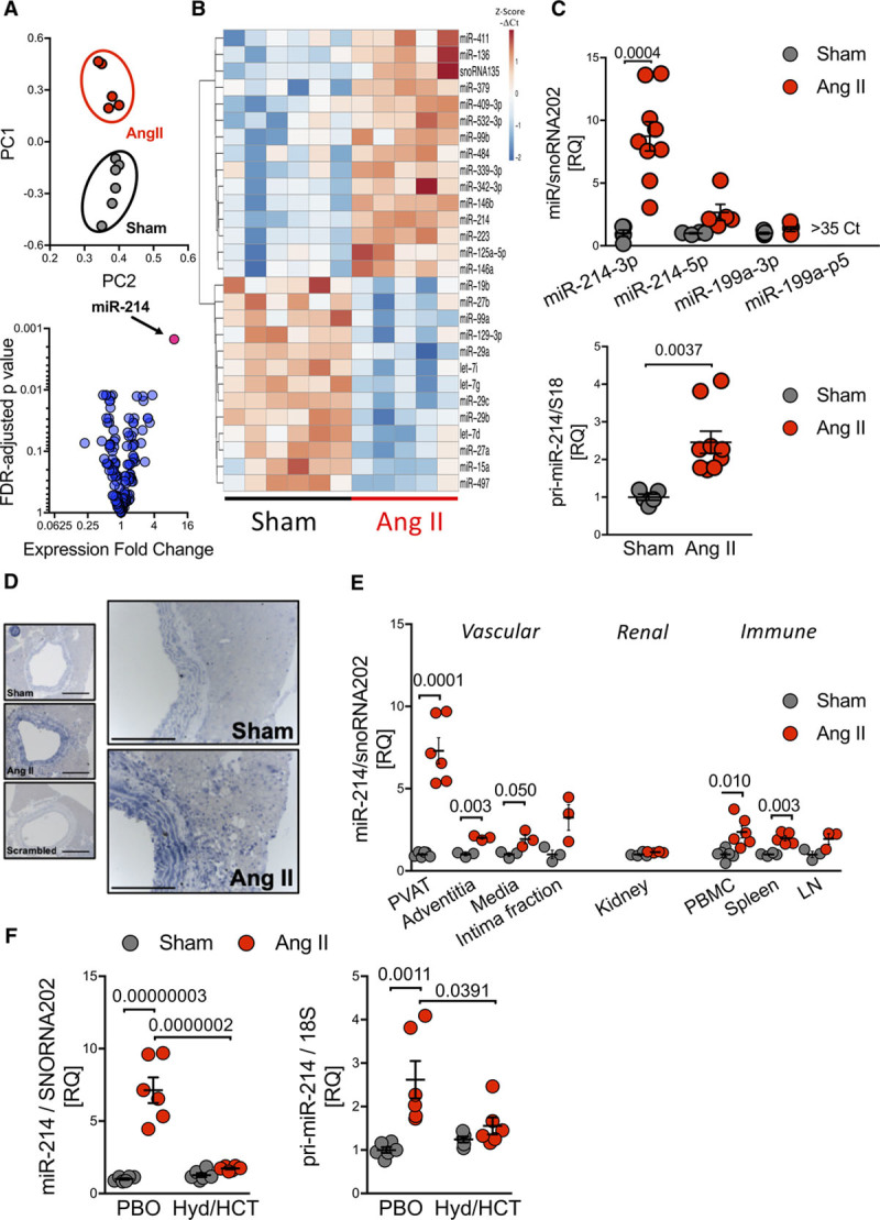
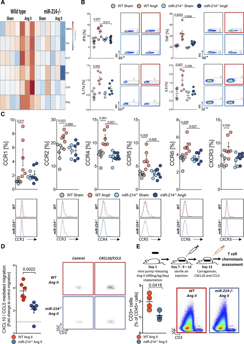
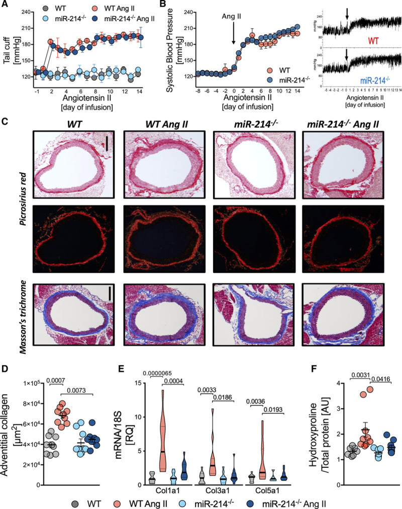
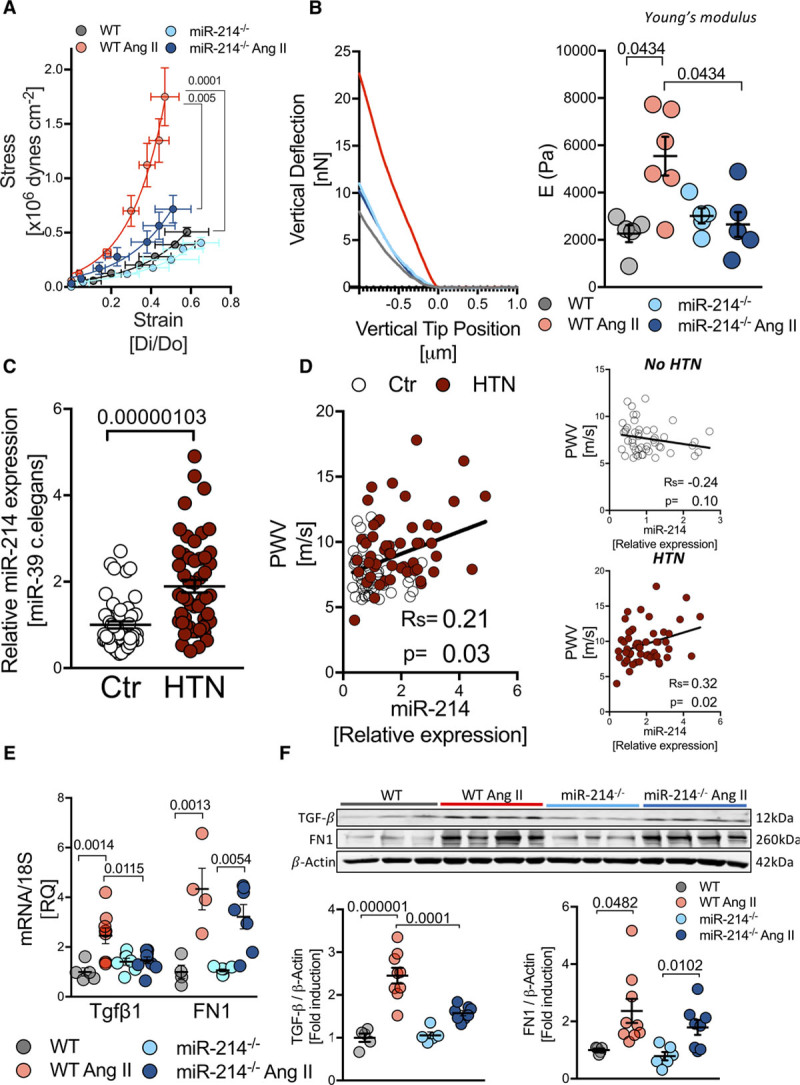
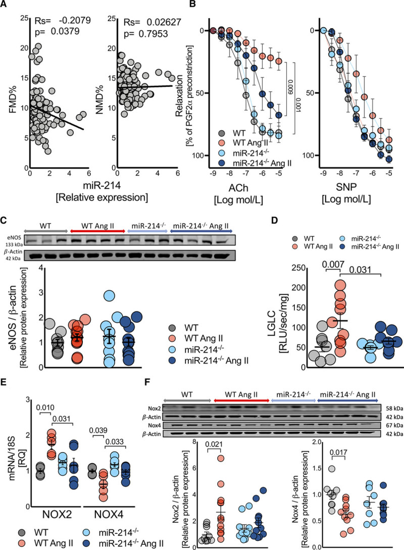
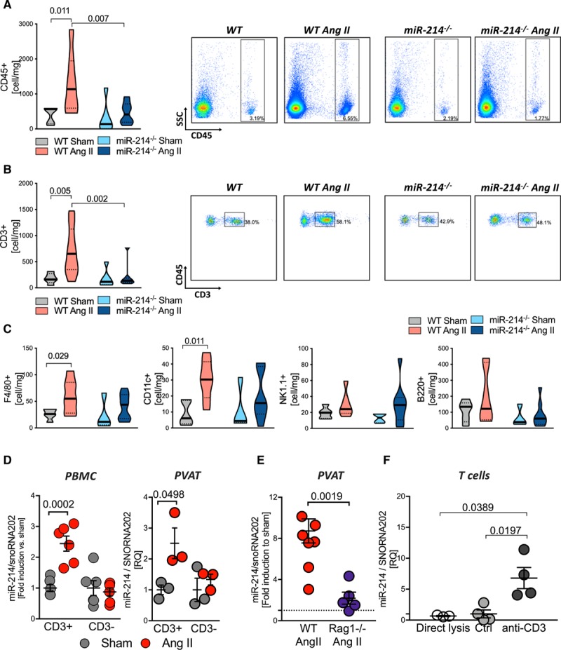
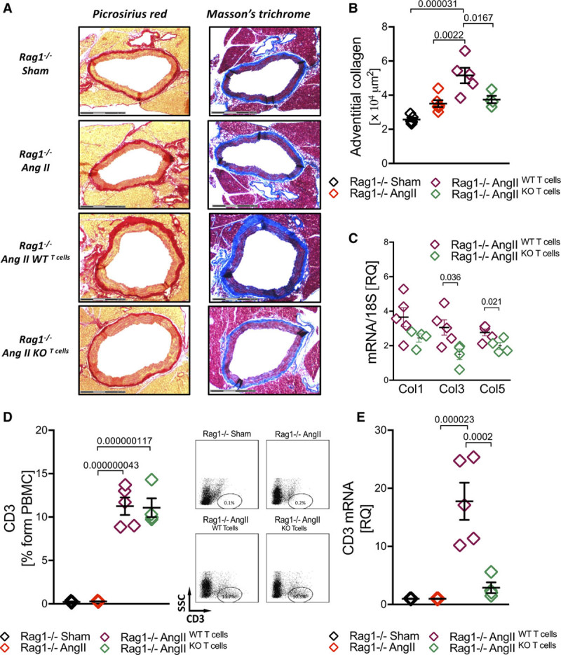
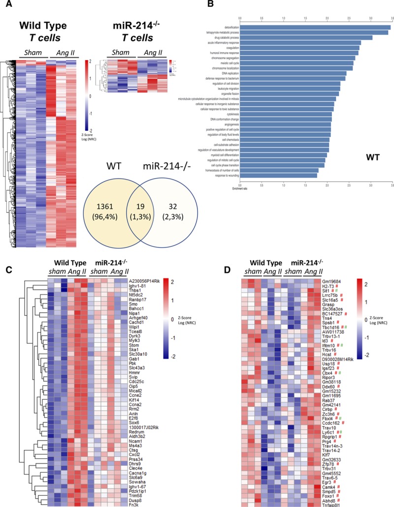
Out of 381 miRs screened in the perivascular tissues in response to Ang II (angiotensin II)-mediated hypertension, miR-214 showed the highest induction (8-fold, =0.0001). MiR-214 induction was pronounced in perivascular and circulating T cells, but not in perivascular adipose tissue adipocytes. Global deletion of miR-214 prevented Ang II-induced periaortic fibrosis, , , and expression, hydroxyproline accumulation, and vascular stiffening, without difference in blood pressure. Mechanistic studies revealed that miR-214 mice were protected against endothelial dysfunction, oxidative stress, and increased Nox2, all of which were induced by Ang II in WT mice. Ang II-induced recruitment of T cells into perivascular adipose tissue was abolished in miR-214 mice. Adoptive transfer of miR-214 T cells into RAG1 mice resulted in reduced perivascular fibrosis compared with the effect of WT T cells. Ang II induced hypertension caused significant change in the expression of 1380 T cell genes in WT, but only 51 in miR-214. T cell activation, proliferation and chemotaxis pathways were differentially affected. MiR-214 prevented Ang II-induction of profibrotic T cell cytokines (, , and ) and chemokine receptors (CCR1, CCR2, CCR4, CCR5, CCR6, and CXCR3). This manifested in reduced in vitro and in vivo T cell chemotaxis resulting in attenuation of profibrotic perivascular inflammation. Translationally, we show that miR-214 is increased in plasma of patients with hypertension and is directly correlated to pulse wave velocity as a measure of vascular stiffness.

CONCLUSIONS

T-cell-derived miR-214 controls pathological perivascular fibrosis in hypertension mediated by T cell recruitment and local profibrotic cytokine release.

摘要

背景

尽管人们越来越了解与高血压相关的血管僵硬与血管周围纤维化有关的预后重要性,但对这一过程的分子和细胞调节仍知之甚少。

目的

研究 microRNA-214(miR-214)在诱导血管周围纤维化和内皮功能障碍导致血管僵硬中的功能作用。

方法和结果

在血管周围组织中,在 Ang II(血管紧张素 II)介导的高血压反应中筛选了 381 种 miRs,miR-214 的诱导最高(8 倍,=0.0001)。miR-214 的诱导在血管周围和循环 T 细胞中很明显,但在血管周围脂肪组织脂肪细胞中则不然。miR-214 的全局缺失可预防 Ang II 诱导的主动脉旁纤维化、、、和表达、羟脯氨酸积累和血管僵硬,而血压无差异。机制研究表明,miR-214 小鼠对内皮功能障碍、氧化应激和 Nox2 增加具有保护作用,所有这些在 WT 小鼠中均由 Ang II 诱导。miR-214 小鼠中 Ang II 诱导的 T 细胞募集到血管周围脂肪组织中被消除。将 miR-214 T 细胞过继转移到 RAG1 小鼠中可减少与 WT T 细胞相比的血管周围纤维化。Ang II 诱导的高血压导致 WT 中 1380 个 T 细胞基因的表达发生显著变化,但 miR-214 中只有 51 个。T 细胞激活、增殖和趋化途径受到不同影响。miR-214 可预防 Ang II 诱导的致纤维化 T 细胞细胞因子(、、和)和趋化因子受体(CCR1、CCR2、CCR4、CCR5、CCR6 和 CXCR3)。这表现在体外和体内 T 细胞趋化作用降低,导致致纤维化的血管周围炎症减轻。在翻译水平上,我们表明 miR-214 在高血压患者的血浆中增加,并与脉搏波速度直接相关,作为血管僵硬的衡量标准。

结论

T 细胞衍生的 miR-214 通过 T 细胞募集和局部致纤维化细胞因子释放控制高血压中的病理性血管周围纤维化。

https://cdn.ncbi.nlm.nih.gov/pmc/blobs/c0e9/7147427/27e30e525700/res-126-988-g008.jpg
https://cdn.ncbi.nlm.nih.gov/pmc/blobs/c0e9/7147427/a28f22882cd3/res-126-988-g001.jpg
https://cdn.ncbi.nlm.nih.gov/pmc/blobs/c0e9/7147427/0203a0eda04e/res-126-988-g002.jpg
https://cdn.ncbi.nlm.nih.gov/pmc/blobs/c0e9/7147427/9ebbe5dc7296/res-126-988-g003.jpg
https://cdn.ncbi.nlm.nih.gov/pmc/blobs/c0e9/7147427/684695b0eaee/res-126-988-g004.jpg
https://cdn.ncbi.nlm.nih.gov/pmc/blobs/c0e9/7147427/aad53333ca1d/res-126-988-g005.jpg
https://cdn.ncbi.nlm.nih.gov/pmc/blobs/c0e9/7147427/1ac1eb7ed16c/res-126-988-g006.jpg
https://cdn.ncbi.nlm.nih.gov/pmc/blobs/c0e9/7147427/39a34452fe39/res-126-988-g007.jpg
https://cdn.ncbi.nlm.nih.gov/pmc/blobs/c0e9/7147427/27e30e525700/res-126-988-g008.jpg

相似文献

1
T-Cell-Derived miRNA-214 Mediates Perivascular Fibrosis in Hypertension.T 细胞来源的 microRNA-214 介导高血压中的血管周围纤维化。
Circ Res. 2020 Apr 10;126(8):988-1003. doi: 10.1161/CIRCRESAHA.119.315428. Epub 2020 Feb 17.
2
Role of chemokine RANTES in the regulation of perivascular inflammation, T-cell accumulation, and vascular dysfunction in hypertension.趋化因子RANTES在高血压中对血管周围炎症、T细胞聚集及血管功能障碍的调节作用
FASEB J. 2016 May;30(5):1987-99. doi: 10.1096/fj.201500088R. Epub 2016 Feb 12.
3
Perivascular Fibrosis Is Mediated by a KLF10-IL-9 Signaling Axis in CD4+ T Cells.血管周纤维化由 CD4+T 细胞中的 KLF10-IL-9 信号轴介导。
Circ Res. 2022 May 27;130(11):1662-1681. doi: 10.1161/CIRCRESAHA.121.320420. Epub 2022 Apr 20.
4
Vascular smooth muscle cell-specific miRNA-214 knockout inhibits angiotensin II-induced hypertension through upregulation of Smad7.血管平滑肌细胞特异性 miRNA-214 敲除通过上调 Smad7 抑制血管紧张素 II 诱导的高血压。
FASEB J. 2021 Nov;35(11):e21947. doi: 10.1096/fj.202100766RR.
5
Endothelial-derived extracellular microRNA-92a promotes arterial stiffness by regulating phenotype changes of vascular smooth muscle cells.内皮细胞衍生的外泌体 microRNA-92a 通过调节血管平滑肌细胞的表型变化促进动脉僵硬。
Sci Rep. 2022 Jan 10;12(1):344. doi: 10.1038/s41598-021-04341-1.
6
MicroRNA-31 Regulates Immunosuppression in Ang II (Angiotensin II)-induced Hypertension by Targeting Ppp6C (Protein Phosphatase 6c).MicroRNA-31 通过靶向 Ppp6C(蛋白磷酸酶 6c)调节 Ang II(血管紧张素 II)诱导的高血压中的免疫抑制。
Hypertension. 2019 May;73(5):e14-e24. doi: 10.1161/HYPERTENSIONAHA.118.12319.
7
γδ T Cells Mediate Angiotensin II-Induced Hypertension and Vascular Injury.γδ T 细胞介导血管紧张素 II 诱导的高血压和血管损伤。
Circulation. 2017 May 30;135(22):2155-2162. doi: 10.1161/CIRCULATIONAHA.116.027058. Epub 2017 Mar 22.
8
Matrix metalloproteinase-2 knockout prevents angiotensin II-induced vascular injury.基质金属蛋白酶-2 敲除可预防血管紧张素 II 诱导的血管损伤。
Cardiovasc Res. 2017 Dec 1;113(14):1753-1762. doi: 10.1093/cvr/cvx115.
9
PTEN (Phosphatase and Tensin Homolog) Protects Against Ang II (Angiotensin II)-Induced Pathological Vascular Fibrosis and Remodeling-Brief Report.PTEN(磷酸酶和张力蛋白同源物)可预防血管紧张素 II(Angiotensin II)诱导的病理性血管纤维化和重塑——简短报告。
Arterioscler Thromb Vasc Biol. 2020 Feb;40(2):394-403. doi: 10.1161/ATVBAHA.119.313757. Epub 2019 Dec 19.
10
SIRT3 (Sirtuin-3) Prevents Ang II (Angiotensin II)-Induced Macrophage Metabolic Switch Improving Perivascular Adipose Tissue Function.SIRT3(沉默调节蛋白 3)可预防血管紧张素 II(血管紧张素 II)诱导的巨噬细胞代谢转换,改善血管周围脂肪组织功能。
Arterioscler Thromb Vasc Biol. 2021 Feb;41(2):714-730. doi: 10.1161/ATVBAHA.120.315337. Epub 2020 Dec 17.

引用本文的文献

1
Arterial stiffness and vascular aging: mechanisms, prevention, and therapy.动脉僵硬度与血管衰老:机制、预防及治疗
Signal Transduct Target Ther. 2025 Sep 1;10(1):282. doi: 10.1038/s41392-025-02346-0.
2
Hepatic stellate cell-specific miR-214 expression alleviates liver fibrosis without boosting steatosis and inflammation.肝星状细胞特异性miR-214表达可减轻肝纤维化,而不会加重脂肪变性和炎症。
J Transl Med. 2025 Jul 22;23(1):810. doi: 10.1186/s12967-025-06880-x.
3
The cross-sectional association between cardiometabolic index and abdominal aortic calcification in U.S. adults: evidence from NHANES 2013-2014.

本文引用的文献

1
Cardiovascular Effects of Pharmacological Targeting of Sphingosine Kinase 1.靶向鞘氨醇激酶 1 的药理学作用对心血管的影响。
Hypertension. 2020 Feb;75(2):383-392. doi: 10.1161/HYPERTENSIONAHA.119.13450. Epub 2019 Dec 16.
2
Causal association between periodontitis and hypertension: evidence from Mendelian randomization and a randomized controlled trial of non-surgical periodontal therapy.牙周炎与高血压的因果关系:来自孟德尔随机化和非手术牙周治疗随机对照试验的证据。
Eur Heart J. 2019 Nov 1;40(42):3459-3470. doi: 10.1093/eurheartj/ehz646.
3
T Cell-Derived IL-17A Induces Vascular Dysfunction via Perivascular Fibrosis Formation and Dysregulation of NO/cGMP Signaling.
美国成年人心脏代谢指数与腹主动脉钙化之间的横断面关联:来自2013 - 2014年美国国家健康与营养检查调查(NHANES)的证据。
Front Nutr. 2025 Jul 2;12:1537795. doi: 10.3389/fnut.2025.1537795. eCollection 2025.
4
[Influence and mechanism of extracellular vesicles derived from human dermal papilla cells on skin fibrosis in mice].[人真皮乳头细胞来源的细胞外囊泡对小鼠皮肤纤维化的影响及机制]
Zhonghua Shao Shang Yu Chuang Mian Xiu Fu Za Zhi. 2025 Jun 20;41(6):559-568. doi: 10.3760/cma.j.cn501225-20240925-00348.
5
Real-world effectiveness and safety of oral azvudine versus Paxlovid in patients with COVID-19 and pre-existing hypertension: a multicentre, retrospective, cohort study in Henan Province, China.口服阿兹夫定与帕罗韦德治疗新型冠状病毒肺炎合并高血压患者的真实世界有效性和安全性:中国河南省一项多中心、回顾性队列研究
BMJ Open. 2025 Jun 27;15(6):e090230. doi: 10.1136/bmjopen-2024-090230.
6
Endothelial MicroRNA-214 Confers Angiotensin II Hypertension by Targeting eNOS in Mice.内皮细胞微小RNA-214通过靶向小鼠内皮型一氧化氮合酶导致血管紧张素II性高血压。
Kidney Blood Press Res. 2025;50(1):442-459. doi: 10.1159/000546674. Epub 2025 May 30.
7
The immune system in cardiovascular diseases: from basic mechanisms to therapeutic implications.心血管疾病中的免疫系统:从基本机制到治疗意义
Signal Transduct Target Ther. 2025 May 23;10(1):166. doi: 10.1038/s41392-025-02220-z.
8
Habitual Exercise Modulates Neuroimmune Interaction to Mitigate Aortic Stiffness.习惯性运动调节神经免疫相互作用以减轻主动脉僵硬。
Circ Res. 2025 Jun 6;136(12):1579-1594. doi: 10.1161/CIRCRESAHA.124.325656. Epub 2025 Apr 30.
9
SPARC: a key mediator of apoptosis in human umbilical vein endothelial cells and its role in hypertension mechanism.SPARC:人脐静脉内皮细胞凋亡的关键介质及其在高血压机制中的作用
In Vitro Cell Dev Biol Anim. 2025 Apr 14. doi: 10.1007/s11626-025-01026-1.
10
Immunotherapy for hypertensive end-organ damage: a new therapeutic strategy.高血压性靶器官损害的免疫治疗:一种新的治疗策略。
Essays Biochem. 2025 Mar 25;0(0):EBC20243000. doi: 10.1042/EBC20243000.
T 细胞衍生的白介素-17A 通过血管周围纤维化形成和一氧化氮/cGMP 信号通路失调诱导血管功能障碍。
Oxid Med Cell Longev. 2019 Jul 18;2019:6721531. doi: 10.1155/2019/6721531. eCollection 2019.
4
Chanzyme TRPM7 protects against cardiovascular inflammation and fibrosis.Chanzyme TRPM7 可预防心血管炎症和纤维化。
Cardiovasc Res. 2020 Mar 1;116(3):721-735. doi: 10.1093/cvr/cvz164.
5
WebGestalt 2019: gene set analysis toolkit with revamped UIs and APIs.WebGestalt 2019:基因集分析工具包,具有全新的用户界面和 API。
Nucleic Acids Res. 2019 Jul 2;47(W1):W199-W205. doi: 10.1093/nar/gkz401.
6
Chemokine Receptor Redundancy and Specificity Are Context Dependent.趋化因子受体的冗余性和特异性是依赖于上下文的。
Immunity. 2019 Feb 19;50(2):378-389.e5. doi: 10.1016/j.immuni.2019.01.009.
7
Monocytes M( MP)aking Way for T-Cell Vascular Infiltration.单核细胞为T细胞血管浸润让路。
Circ Res. 2018 Aug 31;123(6):638-640. doi: 10.1161/CIRCRESAHA.118.313668.
8
A new "lnc" between non-coding RNA and cardiac regeneration.非编码RNA与心脏再生之间的一种新型“长链非编码RNA”
Cardiovasc Res. 2018 Oct 1;114(12):1569-1570. doi: 10.1093/cvr/cvy153.
9
Absence of natriuretic peptide clearance receptor attenuates TGF-β1-induced selective atrial fibrosis and atrial fibrillation.利钠肽清除受体缺失可减轻 TGF-β1 诱导的选择性心房纤维化和心房颤动。
Cardiovasc Res. 2019 Feb 1;115(2):357-372. doi: 10.1093/cvr/cvy224.
10
MicroRNA-214-3p in the Kidney Contributes to the Development of Hypertension.肾中的 microRNA-214-3p 有助于高血压的发展。
J Am Soc Nephrol. 2018 Oct;29(10):2518-2528. doi: 10.1681/ASN.2018020117. Epub 2018 Jul 26.