Department of Radiology, Athinoula A. Martinos Center for Biomedical Imaging, Massachusetts General Hospital, Charlestown, MA, USA.
Harvard Medical School, Boston, MA, USA.
Mol Psychiatry. 2021 May;26(5):1659-1669. doi: 10.1038/s41380-020-0682-z. Epub 2020 Feb 19.
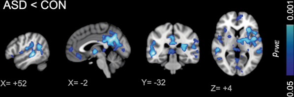
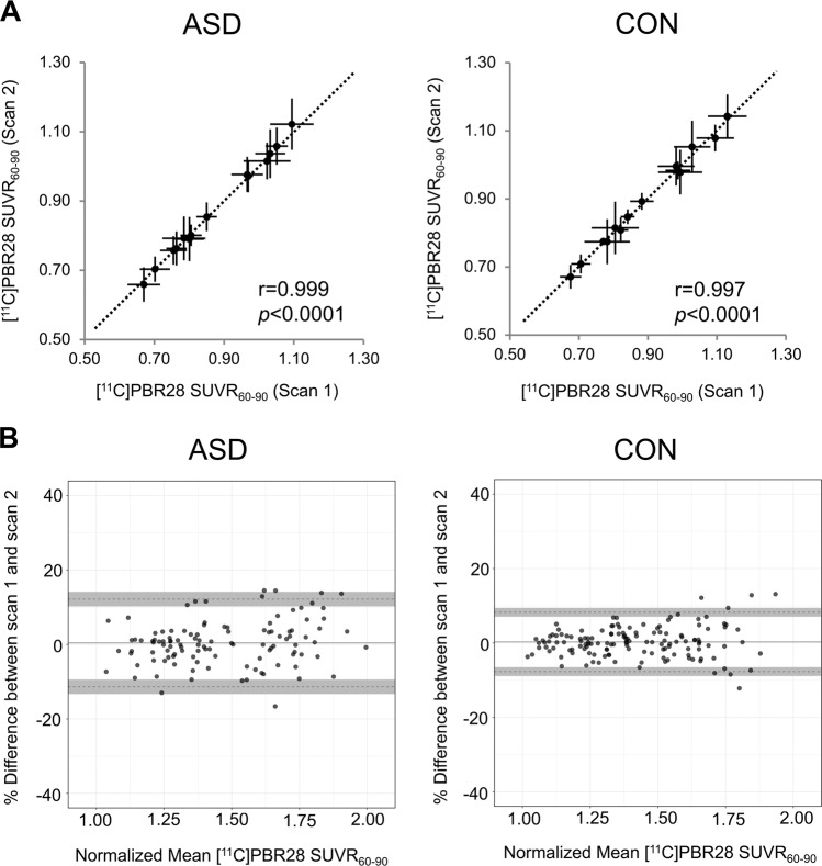
Mechanisms of neuroimmune and mitochondrial dysfunction have been repeatedly implicated in autism spectrum disorder (ASD). To examine these mechanisms in ASD individuals, we measured the in vivo expression of the 18 kDa translocator protein (TSPO), an activated glial marker expressed on mitochondrial membranes. Participants underwent scanning on a simultaneous magnetic resonance-positron emission tomography (MR-PET) scanner with the second-generation TSPO radiotracer [C]PBR28. By comparing TSPO in 15 young adult males with ASD with 18 age- and sex-matched controls, we showed that individuals with ASD exhibited lower regional TSPO expression in several brain regions, including the bilateral insular cortex, bilateral precuneus/posterior cingulate cortex, and bilateral temporal, angular, and supramarginal gyri, which have previously been implicated in autism in functional MR imaging studies. No brain region exhibited higher regional TSPO expression in the ASD group compared with the control group. A subset of participants underwent a second MR-PET scan after a median interscan interval of 3.6 months, and we determined that TSPO expression over this period of time was stable and replicable. Furthermore, voxelwise analysis confirmed lower regional TSPO expression in ASD at this later time point. Lower TSPO expression in ASD could reflect abnormalities in neuroimmune processes or mitochondrial dysfunction.
神经免疫和线粒体功能障碍的机制已被反复牵涉到自闭症谱系障碍(ASD)中。为了在 ASD 个体中研究这些机制,我们测量了活体中 18 kDa 转位蛋白(TSPO)的表达,TSPO 是一种在线粒体膜上表达的活跃神经胶质标志物。参与者在配备有第二代 TSPO 放射性示踪剂 [C]PBR28 的磁共振正电子发射断层扫描(MR-PET)扫描仪上接受扫描。通过将 15 名患有 ASD 的年轻男性与 18 名年龄和性别匹配的对照组的 TSPO 进行比较,我们发现 ASD 个体在几个大脑区域的局部 TSPO 表达较低,包括双侧岛叶、双侧后扣带回/楔前叶和双侧颞叶、角回和缘上回,这些区域在功能磁共振成像研究中与自闭症有关。与对照组相比,ASD 组没有任何大脑区域表现出更高的局部 TSPO 表达。一部分参与者在中位间隔扫描时间为 3.6 个月后进行了第二次 MR-PET 扫描,我们确定在这段时间内 TSPO 的表达是稳定且可重复的。此外,体素分析进一步证实了 ASD 患者在稍后时间点的局部 TSPO 表达降低。ASD 中 TSPO 表达降低可能反映了神经免疫过程或线粒体功能障碍的异常。