Department of Joint Surgery, Third Hospital of Hebei Medical University, Shijiazhuang, Hebei, China.
Department of Orthopaedic Surgery, Affiliated Hospital of Hebei University of Engineering, Handan, Hebei, China.
Orthop Surg. 2020 Apr;12(2):653-660. doi: 10.1111/os.12631. Epub 2020 Feb 19.
To explore the effects of patellectomy on the bony and cartilaginous morphology of the trochlear groove in growing rabbits.
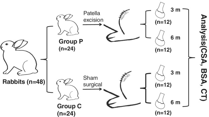
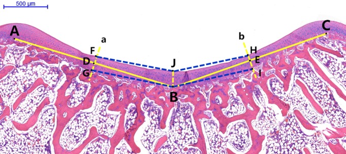
Forty-eight 4-week-old New Zealand white rabbits were randomly assigned to two groups. The control group underwent a sham surgical procedure, whereas the patellectomy group underwent patella excision surgery. Half of the rabbits in each group were sacrificed 3 months postoperatively; the rest were sacrificed 6 months postoperatively. Hematoxylin and eosin staining was performed on collected samples. Measurements included the bony and cartilaginous sulcus angles of the trochlear groove. In addition, the thickness of the articular cartilage at the deepest sulcus position (central thickness) and at the mid-position of the medial and lateral facets was measured and compared between groups.
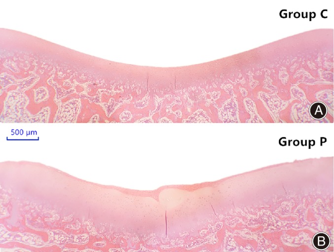
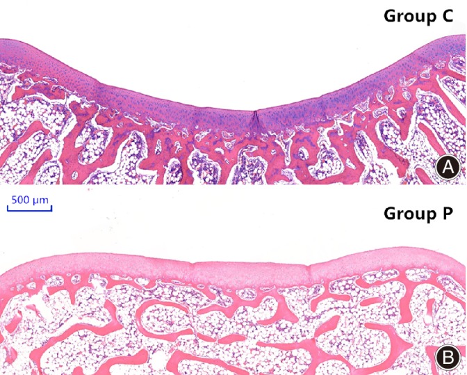
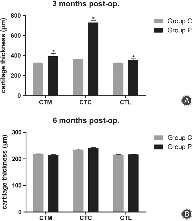
Three months after surgery, histological images revealed significant differences between the control group and the patellectomy group in cartilaginous sulcus angle (144.2° ± 1.5° vs 151.9° ± 2.4°, respectively; P < 0.001). No obvious difference in bony sulcus angle was found between the groups. Six months after surgery, significant between-group differences were observed in cartilaginous sulcus angle (136.3° ± 2.5° in control group vs 160.7° ± 3.0° in patellectomy group, P < 0.001) and bony sulcus angle (136.2° ± 2.2° in control group vs 160.4° ± 2.6° in patellectomy group, P < 0.001). However, there were no significant intra-group differences between cartilaginous and bony sulcus angles in either group. Three months after surgery, significant between-group differences were detected in articular cartilage thickness at the three different positions (medial facet: 324.3 ± 14.0 μm in control group vs 391.7 ± 98.8 μm in patellectomy group, P = 0.029; central position: 362.1 ± 13.6 μm in control group vs 730.3 ± 76.8 μm in patellectomy group, P < 0.001; lateral facet: 324.6 ± 12.7 μm in control group vs 358.5 ± 38.7 μm in patellectomy group, P = 0.009). No between-group differences in cartilage thickness were found at 6 months.
Abnormal mechanical stress (patellectomy) during a rabbit's development can cause flattening of the femoral trochlear cartilage, followed by changes in the subchondral osseous layer. Abnormal mechanical stress is a crucial factor in the development of trochlear groove dysplasia.
探讨髌骨切除对生长兔滑车沟骨软骨形态的影响。
将 48 只 4 周龄新西兰白兔随机分为两组。对照组行假手术,髌骨切除组行髌骨切除术。每组一半的兔子在术后 3 个月处死,其余的在术后 6 个月处死。收集样本后进行苏木精-伊红染色。测量指标包括滑车沟的骨性和软骨性沟角。此外,还测量并比较了各组滑车沟最深位置(中央位置)和内外侧关节面中点处的关节软骨厚度。
术后 3 个月,组织学图像显示对照组和髌骨切除组的软骨沟角有显著差异(分别为 144.2°±1.5°和 151.9°±2.4°;P<0.001)。两组间骨性沟角无明显差异。术后 6 个月,两组间软骨沟角有显著差异(对照组 136.3°±2.5°,髌骨切除组 160.7°±3.0°;P<0.001)和骨性沟角(对照组 136.2°±2.2°,髌骨切除组 160.4°±2.6°;P<0.001)。然而,两组的软骨沟角和骨性沟角均无明显的组内差异。术后 3 个月,在三个不同位置的关节软骨厚度上发现了组间的显著差异(内侧关节面:对照组 324.3±14.0μm,髌骨切除组 391.7±98.8μm,P=0.029);中央位置:对照组 362.1±13.6μm,髌骨切除组 730.3±76.8μm,P<0.001;外侧关节面:对照组 324.6±12.7μm,髌骨切除组 358.5±38.7μm,P=0.009)。6 个月时,两组间软骨厚度无差异。
兔发育过程中异常的机械应力(髌骨切除)可导致股骨滑车软骨变平,随后继发软骨下骨层的变化。异常的机械应力是滑车沟发育不良的关键因素。