Viral Mutation Section, HIV Dynamics and Replication Program, Center for Cancer Research, National Cancer Institute at Frederick, Frederick, MD 21702.
Viral Recombination Section, HIV Dynamics and Replication Program, Center for Cancer Research, National Cancer Institute at Frederick, Frederick, MD 21702.
Proc Natl Acad Sci U S A. 2020 Mar 10;117(10):5486-5493. doi: 10.1073/pnas.1920631117. Epub 2020 Feb 24.
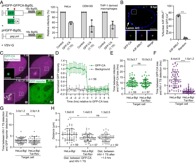
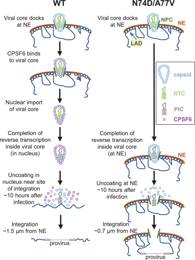
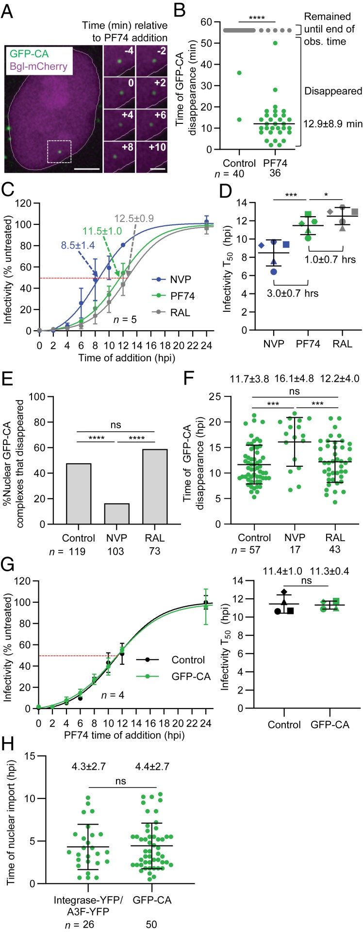
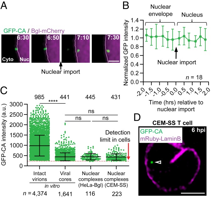
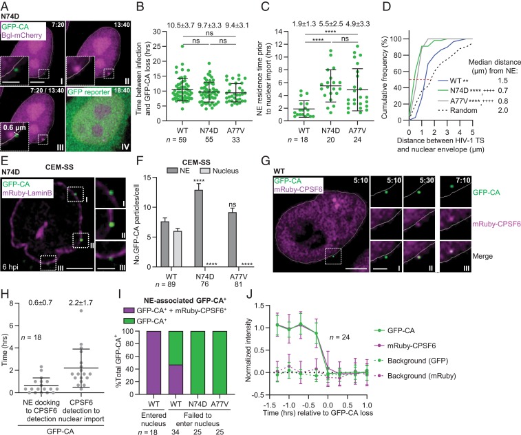
HIV-1 capsid core disassembly (uncoating) must occur before integration of viral genomic DNA into the host chromosomes, yet remarkably, the timing and cellular location of uncoating is unknown. Previous studies have proposed that intact viral cores are too large to fit through nuclear pores and uncoating occurs in the cytoplasm in coordination with reverse transcription or at the nuclear envelope during nuclear import. The capsid protein (CA) content of the infectious viral cores is not well defined because methods for directly labeling and quantifying the CA in viral cores have been unavailable. In addition, it has been difficult to identify the infectious virions because only one of ∼50 virions in infected cells leads to productive infection. Here, we developed methods to analyze HIV-1 uncoating by direct labeling of CA with GFP and to identify infectious virions by tracking viral cores in living infected cells through viral DNA integration and proviral DNA transcription. Astonishingly, our results show that intact (or nearly intact) viral cores enter the nucleus through a mechanism involving interactions with host protein cleavage and polyadenylation specificity factor 6 (CPSF6), complete reverse transcription in the nucleus before uncoating, and uncoat <1.5 h before integration near (<1.5 μm) their genomic integration sites. These results fundamentally change our current understanding of HIV-1 postentry replication events including mechanisms of nuclear import, uncoating, reverse transcription, integration, and evasion of innate immunity.
HIV-1 衣壳核心的解体(脱壳)必须发生在病毒基因组 DNA 整合到宿主染色体之前,但令人惊讶的是,脱壳的时间和细胞位置尚不清楚。先前的研究提出,完整的病毒核心太大,无法通过核孔,脱壳发生在细胞质中,与逆转录协调进行,或在核输入过程中发生在核膜上。感染性病毒核心的衣壳蛋白 (CA) 含量尚未得到很好的定义,因为直接标记和定量病毒核心中的 CA 的方法尚未出现。此外,由于只有感染细胞中约 50 个病毒粒子中的一个能够导致有效感染,因此很难识别感染性病毒粒子。在这里,我们开发了通过 GFP 直接标记 CA 来分析 HIV-1 脱壳的方法,并通过跟踪感染细胞中活病毒核心的病毒 DNA 整合和前病毒 DNA 转录来识别感染性病毒粒子。令人惊讶的是,我们的结果表明,完整的(或几乎完整的)病毒核心通过一种涉及与宿主蛋白切割和多聚腺苷酸化特异性因子 6(CPSF6)相互作用的机制进入细胞核,在脱壳之前在细胞核中完成逆转录,然后在整合到基因组整合位点附近 (<1.5 μm) 之前约 1.5 小时脱壳。这些结果从根本上改变了我们对 HIV-1 进入后复制事件的现有理解,包括核输入、脱壳、逆转录、整合和逃避先天免疫的机制。