Alvarez Alyssa, Youn Gun Min, Remigio Van Gogh Angelica Marie, Shin Yin Sophia Sarang, Chakrabarti Moyukh O, McGahan Patrick J, Chen James L
Advanced Orthopaedics and Sports Medicine, San Francisco, California, U.S.A.
Arthrosc Tech. 2020 Jan 8;9(2):e213-e216. doi: 10.1016/j.eats.2019.09.023. eCollection 2020 Feb.
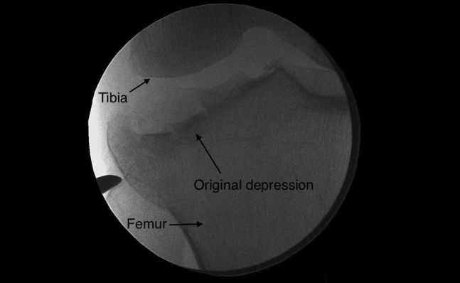
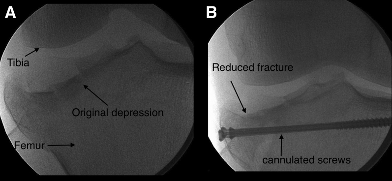
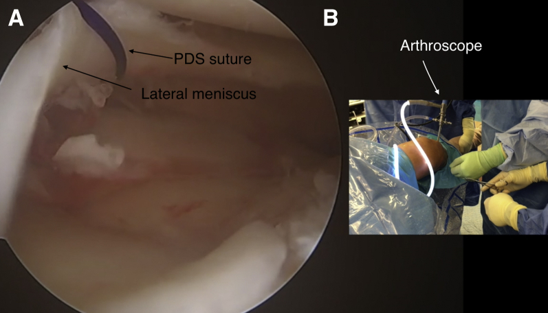
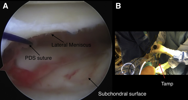
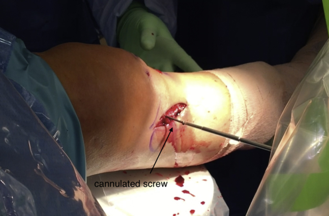
Arthroscopic-assisted internal fixation is an ideal technique for visualizing chondral reduction during tibial open reduction-internal fixation. Typically, open reduction-internal fixation is performed using radiographic and Fluoroscan imaging (Hologic, Bedford, MA) for reduction of subchondral bone. However, reduction without visualization does not ensure chondral surface reduction. This Technical Note and supplemental video describe an arthroscopic-assisted technique involving the tibial plateau that gives complete visualization as tamping occurs to restore the cartilage surface of the subchondral bone and elevate the fracture.
关节镜辅助下内固定是在胫骨切开复位内固定术中可视化软骨复位的理想技术。通常,切开复位内固定术是使用X线摄影和Fluoroscan成像(Hologic,贝德福德,马萨诸塞州)来复位软骨下骨。然而,在没有可视化的情况下进行复位并不能确保软骨表面的复位。本技术说明及补充视频介绍了一种涉及胫骨平台的关节镜辅助技术,该技术在夯实过程中能提供完整的可视化,以恢复软骨下骨的软骨表面并抬高骨折部位。