Libonati Antonio, Di Taranto Virginia, Gallusi Gianni, Montemurro Edoardo, Campanella Vincenzo
Department of Clinical Sciences and Translational Medicine, University Tor Vergata, Rome, Italy.
Clin Cosmet Investig Dent. 2020 Feb 10;12:17-24. doi: 10.2147/CCIDE.S237442. eCollection 2020.
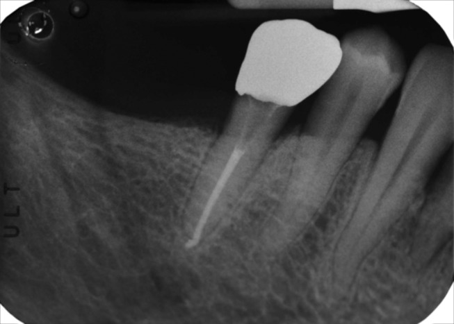
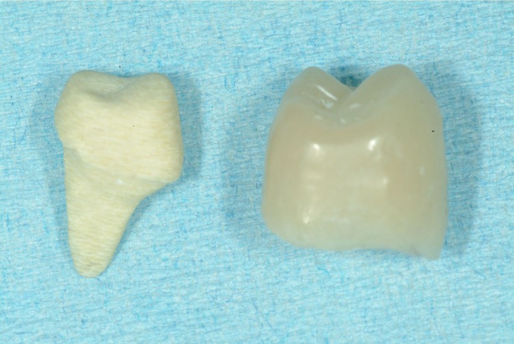
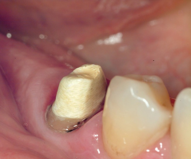
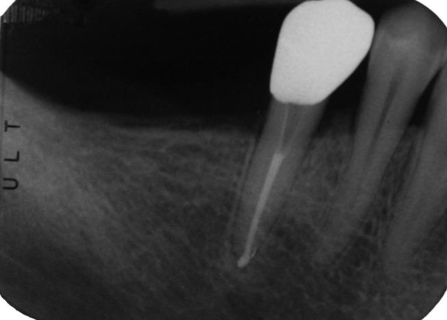
After endodontic treatment, a proper restorative technique is necessary to ensure coronal seal and protection of residual dental structure; teeth which have lost two or more walls need to be restored with posts to increase retention and stability of final restoration. Posts can be distinguished in prefabricated and customized, which are manufactured by lost wax technique or CAD-CAM.
Digital dentistry has been developed to increase workflow precision and to accelerate production process; use of CAD-CAM to realize customized posts was limited to scanning plaster models obtained from traditional impressions.
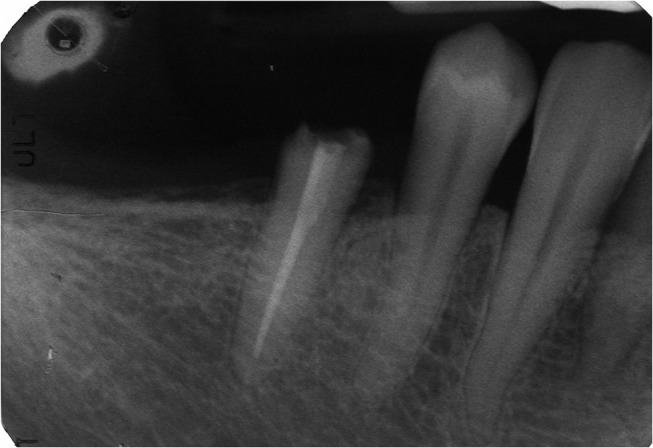
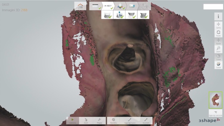
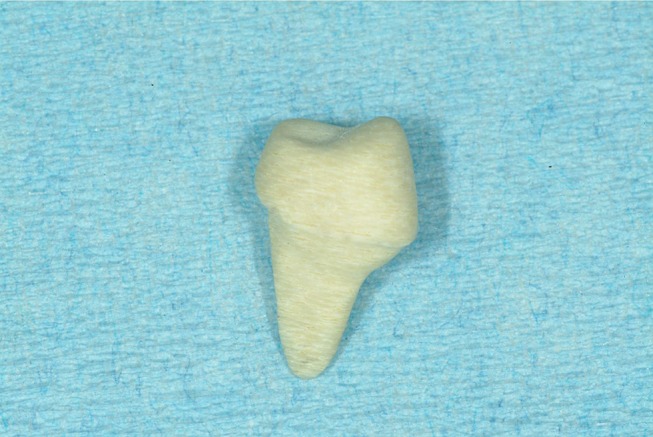
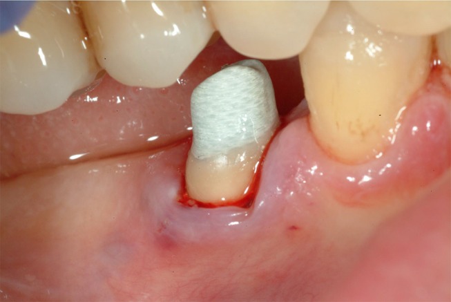
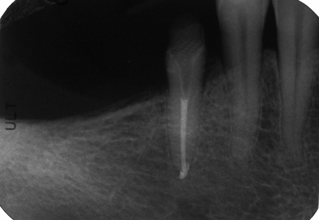
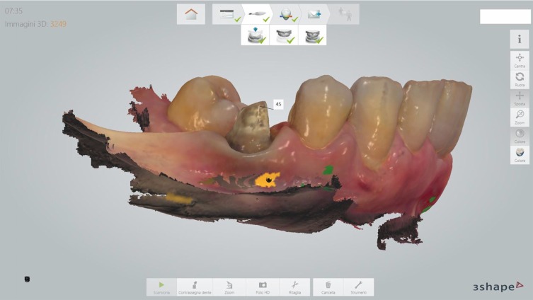
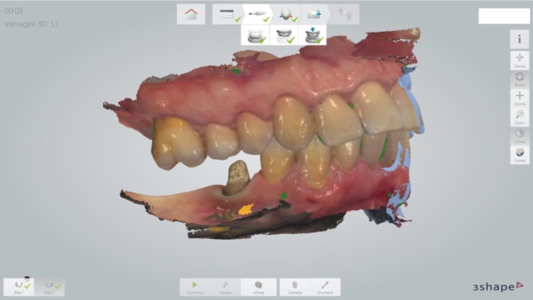
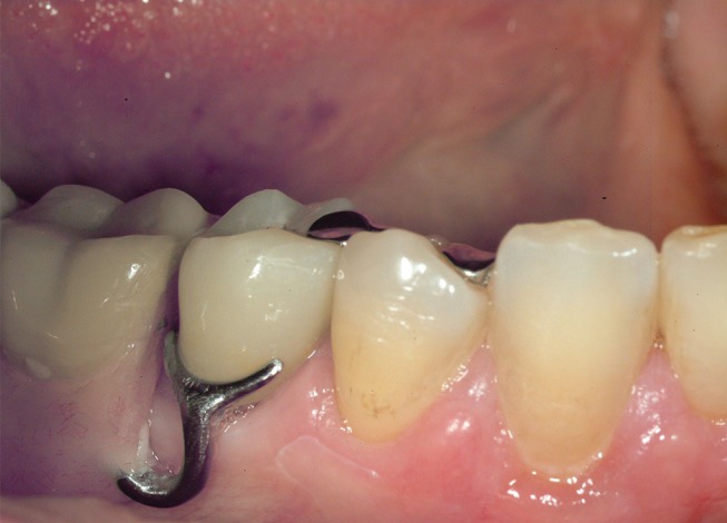
In the reported case an intraoral scan was used to mill a post and core restoration on an endodontically treated inferior premolar; this operative protocol was based on previous in vitro experiments that confirmed the ability of 3shape Trios scanner to read post-space up to 9 mm in depth.
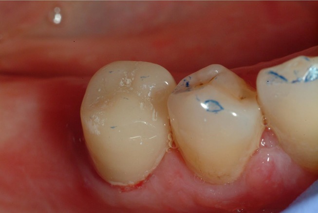
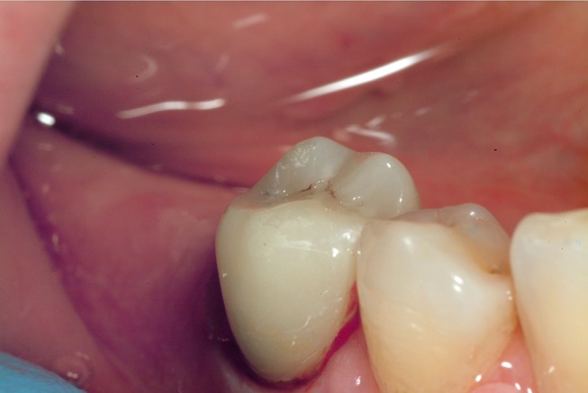
The digital technique allows us to convert the concave surface of the root canal into the convex surface of the post, and realize an anatomical post and core that improves the biomechanics of the endodontically treated tooth reducing the possibility of root fractures.
The use of an intraoral digital scanner represents an opportunity for the clinician as it speeds up the production of an anatomical post and core restorations.
根管治疗后,需要采用合适的修复技术来确保冠部封闭并保护剩余牙体结构;失去两个或更多牙壁的牙齿需要用桩修复,以增加最终修复体的固位力和稳定性。桩可分为预成桩和定制桩,后者通过失蜡法或计算机辅助设计与制造(CAD-CAM)技术制作。
数字牙科技术的发展旨在提高工作流程的精确性并加速生产过程;使用CAD-CAM制作定制桩仅限于扫描从传统印模获得的石膏模型。
在本报告病例中,使用口内扫描仪为一颗根管治疗后的下颌前磨牙制作桩核修复体;该手术方案基于先前的体外实验,该实验证实了3shape Trios扫描仪能够读取深度达9毫米的桩道空间。
数字技术使我们能够将根管的凹面转化为桩的凸面,并制作出解剖形态的桩核,改善根管治疗后牙齿的生物力学性能,降低牙根折断的可能性。
使用口内数字扫描仪为临床医生提供了一个机会,因为它加快了解剖形态桩核修复体的制作。