• 文献检索
  • 文档翻译
  • 深度研究
  • 学术资讯
  • Suppr Zotero 插件Zotero 插件
  • 邀请有礼
  • 套餐&价格
  • 历史记录
应用&插件
Suppr Zotero 插件Zotero 插件浏览器插件Mac 客户端Windows 客户端微信小程序
定价
高级版会员购买积分包购买API积分包
服务
文献检索文档翻译深度研究API 文档MCP 服务
关于我们
关于 Suppr公司介绍联系我们用户协议隐私条款
关注我们

Suppr 超能文献

核心技术专利:CN118964589B侵权必究
粤ICP备2023148730 号-1Suppr @ 2026

文献检索

告别复杂PubMed语法,用中文像聊天一样搜索,搜遍4000万医学文献。AI智能推荐,让科研检索更轻松。

立即免费搜索

文件翻译

保留排版,准确专业,支持PDF/Word/PPT等文件格式,支持 12+语言互译。

免费翻译文档

深度研究

AI帮你快速写综述,25分钟生成高质量综述,智能提取关键信息,辅助科研写作。

立即免费体验

黑素细胞病变皮肤镜下的微斑与黑色素瘤相关:一项横断面研究。

Microblotches on Dermoscopy of Melanocytic Lesions are Associated with Melanoma: A Cross-sectional Study.

机构信息

Department of Dermatology, Melanoma Unit, Hospital Clínic of Barcelona, Barcelona, Spain.

出版信息

Acta Derm Venereol. 2020 Apr 6;100(8):adv00106. doi: 10.2340/00015555-3436.

DOI:10.2340/00015555-3436
PMID:32110813
原文链接:https://pmc.ncbi.nlm.nih.gov/articles/PMC9128959/
Abstract

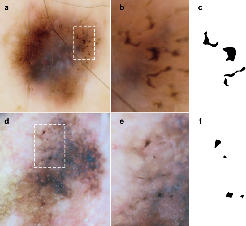
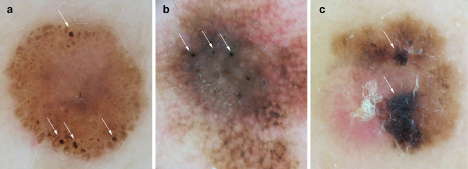
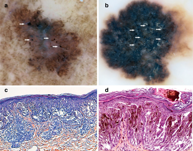
Numerous dermoscopic structures for the early detection of melanoma have been described. The aim of this study was to illustrate the characteristics of dermoscopic structures that are similar to blotches, but smaller (termed microblotches), and to evaluate their association with other well-known dermoscopic structures. A cross-sectional study design, including 165 dermoscopic images of melanoma was used to define microblotches, and 241 consecutive images of naevi from the HAM10000 database, were studied to evaluate the prevalence of this criterion in both groups. Microblotches were defined as sharply demarcated structures ≤1 mm, with geographical borders visible only with dermoscopy. Microblotches were present in 38.7% of the melanomas and 6.7% of the naevi. Moreover, microblotches were associated with an odds ratio (OR) of malignancy of 5.79, and were more frequent in invasive melanoma than in the in-situ subtype (OR 2.92). Histologically, they correspond to hyperpigmented parakeratosis or epidermal consumption. In conclusion, microblotches are related to melanomas. This finding could help dermatologists to differentiate between naevi and melanomas.

摘要

已经描述了许多用于早期检测黑色素瘤的皮肤镜结构。本研究的目的是说明与斑类似但较小(称为微斑)的皮肤镜结构的特征,并评估它们与其他著名的皮肤镜结构的关系。本研究采用了一项横断面研究设计,纳入了 165 张黑色素瘤的皮肤镜图像来定义微斑,并研究了 HAM10000 数据库中 241 张连续的痣的图像,以评估该标准在两组中的发生率。微斑被定义为界限清晰的结构,大小≤1 毫米,仅通过皮肤镜才能看到地理边界。微斑在 38.7%的黑色素瘤和 6.7%的痣中存在。此外,微斑的恶性肿瘤比值比(OR)为 5.79,在侵袭性黑色素瘤中比原位亚型更常见(OR 2.92)。组织学上,它们对应于过度色素沉着角化不全或表皮消耗。总之,微斑与黑色素瘤有关。这一发现可以帮助皮肤科医生区分痣和黑色素瘤。

https://cdn.ncbi.nlm.nih.gov/pmc/blobs/1c1f/9128959/114e656766c4/ActaDV-100-8-5696-g003.jpg
https://cdn.ncbi.nlm.nih.gov/pmc/blobs/1c1f/9128959/4efdc07e179a/ActaDV-100-8-5696-g001.jpg
https://cdn.ncbi.nlm.nih.gov/pmc/blobs/1c1f/9128959/c5ffb5099d6e/ActaDV-100-8-5696-g002.jpg
https://cdn.ncbi.nlm.nih.gov/pmc/blobs/1c1f/9128959/114e656766c4/ActaDV-100-8-5696-g003.jpg
https://cdn.ncbi.nlm.nih.gov/pmc/blobs/1c1f/9128959/4efdc07e179a/ActaDV-100-8-5696-g001.jpg
https://cdn.ncbi.nlm.nih.gov/pmc/blobs/1c1f/9128959/c5ffb5099d6e/ActaDV-100-8-5696-g002.jpg
https://cdn.ncbi.nlm.nih.gov/pmc/blobs/1c1f/9128959/114e656766c4/ActaDV-100-8-5696-g003.jpg

相似文献

1
Microblotches on Dermoscopy of Melanocytic Lesions are Associated with Melanoma: A Cross-sectional Study.黑素细胞病变皮肤镜下的微斑与黑色素瘤相关:一项横断面研究。
Acta Derm Venereol. 2020 Apr 6;100(8):adv00106. doi: 10.2340/00015555-3436.
2
Neck Melanoma: Clinical, Dermoscopic and Confocal Features.颈部黑素瘤:临床、皮肤镜和共聚焦特征。
Dermatology. 2020;236(3):241-247. doi: 10.1159/000503284. Epub 2019 Nov 8.
3
Clinical and Histopathologic Characteristics of Melanocytic Lesions on the Volar Skin Without Typical Dermoscopic Patterns.掌跖部无典型皮肤镜特征的黑素细胞病变的临床及组织病理学特征。
JAMA Dermatol. 2019 May 1;155(5):578-584. doi: 10.1001/jamadermatol.2018.5926.
4
Cutaneous Melanoma Arising in Congenital Melanocytic Nevus: A Retrospective Observational Study.先天性黑素细胞痣中发生的皮肤黑素瘤:一项回顾性观察研究。
Dermatology. 2021;237(3):473-478. doi: 10.1159/000510221. Epub 2020 Oct 14.
5
In vivo confocal microscopy: The role of comparative approach in patients with multiple atypical nevi.体内共聚焦显微镜:比较方法在多发性非典型痣患者中的作用。
Exp Dermatol. 2020 Oct;29(10):945-952. doi: 10.1111/exd.14162. Epub 2020 Sep 21.
6
Association between the dermoscopic morphology of peripheral globules and melanocytic lesion diagnosis.周围小球体的皮肤镜形态与黑素细胞病变诊断的相关性。
J Eur Acad Dermatol Venereol. 2021 Apr;35(4):892-899. doi: 10.1111/jdv.17035. Epub 2020 Dec 23.
7
Digital dermoscopic changes during follow-up of de-novo and nevus-associated melanoma: a cohort study.新发和痣相关黑色素瘤随访中的数字皮肤镜变化:一项队列研究。
Int J Dermatol. 2020 Jul;59(7):813-821. doi: 10.1111/ijd.14918. Epub 2020 May 13.
8
Analysis of the dermoscopic features of excised melanomas and their relation with tumor thickness in a tertiary hospital in Brazil.巴西一家三级医院切除黑色素瘤的皮肤镜特征分析及其与肿瘤厚度的关系。
Int J Dermatol. 2024 Aug;63(8):1064-1070. doi: 10.1111/ijd.17047. Epub 2024 Feb 28.
9
Comparative dermoscopy assessment of nevus-associated versus de novo in situ melanoma.痣相关原位黑色素瘤与新发原位黑色素瘤的对比皮肤镜评估。
Eur J Dermatol. 2023 Apr 1;33(2):121-125. doi: 10.1684/ejd.2023.4454.
10
Digital dermoscopy monitoring of melanocytic lesions: Two novel calculators combining static and dynamic features to identify melanoma.数字皮肤镜监测黑色素瘤病变:两种新型计算器,结合静态和动态特征,以识别黑色素瘤。
J Eur Acad Dermatol Venereol. 2022 Mar;36(3):391-402. doi: 10.1111/jdv.17852. Epub 2021 Dec 17.

引用本文的文献

1
Proposal of a new dermoscopic criterion for pigmented basal cell carcinoma: a multicentre retrospective study.色素性基底细胞癌新的皮肤镜诊断标准的提议:一项多中心回顾性研究
Dermatol Reports. 2023 Aug 24;16(1):9691. doi: 10.4081/dr.2023.9691. eCollection 2024 Mar 12.
2
Frequency of Publication of Dermoscopic Images in Inter-observer Studies: A Systematic Review.观察者间研究中皮肤镜图像发表频率的系统评价。
Acta Derm Venereol. 2021 Dec 17;101(12):adv00621. doi: 10.2340/actadv.v101.865.

本文引用的文献

1
The HAM10000 dataset, a large collection of multi-source dermatoscopic images of common pigmented skin lesions.HAM10000 数据集,一个大型的常见色素性皮肤病变多源皮肤镜图像集合。
Sci Data. 2018 Aug 14;5:180161. doi: 10.1038/sdata.2018.161.
2
Recognition of early melanoma: a monocentric dermoscopy follow-up study comparing de novo melanoma with nevus-associated melanoma.早期黑色素瘤的识别:一项比较新发黑色素瘤与痣相关黑色素瘤的单中心皮肤镜随访研究。
Int J Dermatol. 2018 Jun;57(6):692-702. doi: 10.1111/ijd.13977. Epub 2018 Apr 2.
3
Dermoscopic characteristics of melanoma according to the criteria "ulceration" and "mitotic rate" of the AJCC 2009 staging system for melanoma.
根据美国癌症联合委员会(AJCC)2009年黑色素瘤分期系统的“溃疡形成”和“有丝分裂率”标准,黑色素瘤的皮肤镜特征。
PLoS One. 2017 Apr 11;12(4):e0174871. doi: 10.1371/journal.pone.0174871. eCollection 2017.
4
Dermoscopic features predicting the presence of mitoses in thin melanoma.预测薄型黑色素瘤中存在有丝分裂的皮肤镜特征。
J Dermatol Sci. 2017 May;86(2):158-161. doi: 10.1016/j.jdermsci.2017.01.013. Epub 2017 Feb 3.
5
Standardization of terminology in dermoscopy/dermatoscopy: Results of the third consensus conference of the International Society of Dermoscopy.皮肤镜检查术语的标准化:国际皮肤镜学会第三次共识会议结果
J Am Acad Dermatol. 2016 Jun;74(6):1093-106. doi: 10.1016/j.jaad.2015.12.038. Epub 2016 Feb 17.
6
Dermoscopy structures as predictors of sentinel lymph node positivity in cutaneous melanoma.皮肤镜结构预测皮肤黑色素瘤前哨淋巴结阳性。
Br J Dermatol. 2015;172(5):1269-77. doi: 10.1111/bjd.13552. Epub 2015 Apr 1.
7
Dermoscopy of Naevus-associated Melanomas.痣相关黑色素瘤的皮肤镜检查
Acta Derm Venereol. 2015 Jul;95(6):671-5. doi: 10.2340/00015555-2009.
8
Shiny white streaks: a sign of malignancy at dermoscopy of pigmented skin lesions.亮白色条纹:色素性皮肤病变皮肤镜检查中的恶性征象。
Acta Derm Venereol. 2014 Mar;94(2):132-7. doi: 10.2340/00015555-1683.
9
The impact of dermoscopy on the management of pigmented lesions in everyday clinical practice of general dermatologists: a prospective study.皮肤镜检查对皮肤科医生日常临床实践中色素性皮损处理的影响:一项前瞻性研究。
Br J Dermatol. 2010 Mar;162(3):563-7. doi: 10.1111/j.1365-2133.2009.09551.x. Epub 2009 Oct 15.
10
Dermoscopy compared with naked eye examination for the diagnosis of primary melanoma: a meta-analysis of studies performed in a clinical setting.皮肤镜与肉眼检查在原发性黑色素瘤诊断中的比较:在临床环境中进行的研究的荟萃分析。
Br J Dermatol. 2008 Sep;159(3):669-76. doi: 10.1111/j.1365-2133.2008.08713.x. Epub 2008 Jul 4.