Derwael Céline, Lavergne Olivier, Lovinfosse Pierre, Nechifor Vlad, Salvé Mallory, Waltregny David, Hustinx Roland, Withofs Nadia
Division of Nuclear Medicine and Oncological Imaging, Department of Medical Physics, CHU of Liege, Avenue de l'Hôpital, 1, 4000, Liege, Belgium.
Department of Urology, CHR of Liege, Liege, Belgium.
EJNMMI Res. 2020 Feb 28;10(1):15. doi: 10.1186/s13550-020-0596-4.
Prostate-specific membrane antigen (PSMA) ligand PET/CT has already provided promising results in prostate cancer (PC) imaging, yet simple and reproductible reporting criteria are still lacking. This study aimed at retrospectively evaluating interobserver agreement of [Ga]Ga-PSMA-11 PET/CT images interpretation according to PC molecular imaging standardized evaluation (PROMISE) criteria and reproducibility of PSMA reporting and data systems (RADS).
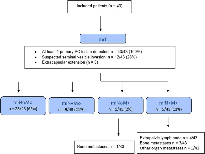
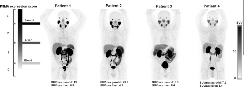
Forty-three patients with newly diagnosed, histologically proven intermediate- or high-risk PC, eligible for radical prostatectomy and who underwent [Ga]Ga-PSMA-11 PET/CT before surgery were retrospectively included. Three nuclear medicine physicians (2 experienced and 1 resident) independently reviewed PET/CT images. Interpretation of [Ga]Ga-PSMA-11 PET/CT images was based on PROMISE criteria including miTNM staging and lesions miPSMA expression score visual estimation and PSMA-RADS version 1.0 for a given scan. Readers' agreement was measured using Krippendorff's coefficients RESULTS: Agreement between observers was almost perfect (coefficient ≥ 0.81) for miM; it was substantial (coefficient ≥ 0.61) for the following criteria: miT, miN, PSMA-RADS, and miPSMA expression score of primary PC lesion and metastases. However, agreement was moderate (coefficient = 0.41-0.60) for miPSMA score of positive lymph nodes and for detection of PC primary lesion.
Visual interpretation of [Ga]Ga-PSMA-11 PET/CT images in patients with newly diagnosed PC in a clinical setting leads to at least substantial agreement for PROMISE criteria and PSMA-RADS classification except for PC primary lesion detection and for miPSMA expression scoring of positive lymph nodes that might have been hampered by the interindividual variability of reference organs PSMA expression.
前列腺特异性膜抗原(PSMA)配体PET/CT在前列腺癌(PC)成像中已取得了有前景的结果,但仍缺乏简单且可重复的报告标准。本研究旨在根据PC分子成像标准化评估(PROMISE)标准回顾性评估[镓]镓-PSMA-11 PET/CT图像解读的观察者间一致性以及PSMA报告和数据系统(RADS)的可重复性。
回顾性纳入43例新诊断的、经组织学证实为中高危PC且适合行根治性前列腺切除术并在手术前行[镓]镓-PSMA-11 PET/CT检查的患者。三名核医学医师(2名经验丰富的和1名住院医师)独立阅片。[镓]镓-PSMA-11 PET/CT图像解读基于PROMISE标准,包括miTNM分期和病变miPSMA表达评分视觉估计以及针对给定扫描的PSMA-RADS 1.0版标准。使用Krippendorff系数测量读者间的一致性。结果:观察者间对于miM的一致性几乎为完美(系数≥0.81);对于以下标准,一致性为实质性(系数≥0.61):miT、miN、PSMA-RADS以及原发性PC病变和转移灶的miPSMA表达评分。然而,对于阳性淋巴结的miPSMA评分以及PC原发性病变的检测,一致性为中等(系数 = 0.41 - 0.60)。
在临床环境中,对新诊断PC患者的[镓]镓-PSMA-11 PET/CT图像进行视觉解读,除了PC原发性病变检测以及阳性淋巴结的miPSMA表达评分可能因参考器官PSMA表达的个体间变异性而受到影响外,对于PROMISE标准和PSMA-RADS分类至少达成了实质性一致。