• 文献检索
  • 文档翻译
  • 深度研究
  • 学术资讯
  • Suppr Zotero 插件Zotero 插件
  • 邀请有礼
  • 套餐&价格
  • 历史记录
应用&插件
Suppr Zotero 插件Zotero 插件浏览器插件Mac 客户端Windows 客户端微信小程序
定价
高级版会员购买积分包购买API积分包
服务
文献检索文档翻译深度研究API 文档MCP 服务
关于我们
关于 Suppr公司介绍联系我们用户协议隐私条款
关注我们

Suppr 超能文献

核心技术专利:CN118964589B侵权必究
粤ICP备2023148730 号-1Suppr @ 2026

文献检索

告别复杂PubMed语法,用中文像聊天一样搜索,搜遍4000万医学文献。AI智能推荐,让科研检索更轻松。

立即免费搜索

文件翻译

保留排版,准确专业,支持PDF/Word/PPT等文件格式,支持 12+语言互译。

免费翻译文档

深度研究

AI帮你快速写综述,25分钟生成高质量综述,智能提取关键信息,辅助科研写作。

立即免费体验

术前PSMA成像分期中骨显像结果不明确的意义:PSMA-RADS 1.0版的验证

The significance of equivocal bone findings in staging PSMA imaging in the preoperative setting: validation of the PSMA-RADS version 1.0.

作者信息

Kuten Jonathan, Dekalo Snir, Mintz Ishai, Yossepowitch Ofer, Mano Roy, Even-Sapir Einat

机构信息

Departments of Nuclear Medicine, Tel-Aviv Sourasky Medical Center, 6 Weizmann St, 6423906, Tel-Aviv, Israel.

Sackler School of Medicine, Tel-Aviv University, Tel-Aviv, Israel.

出版信息

EJNMMI Res. 2021 Jan 6;11(1):3. doi: 10.1186/s13550-020-00745-8.

DOI:10.1186/s13550-020-00745-8
PMID:33409930
原文链接:https://pmc.ncbi.nlm.nih.gov/articles/PMC7788112/
Abstract

BACKGROUND

Assessing the extent of disease in newly diagnosed prostate cancer (PC) patients is crucial for tailoring an appropriate treatment approach. Prostate-specific membrane antigen (PSMA)-targeted positron emission tomography/computed tomography (PET/CT) reportedly has greater accuracy than conventional imaging for staging PC. As with any imaging modality, pitfalls and nonspecific findings do occur. The PSMA reporting and data system (PSMA-RADS) version 1.0 offers structured interpretation of PSMA-targeted studies and classifies lesions by likelihood of clinical significance. The aim of this retrospective study was to evaluate the clinical significance of equivocal bone findings on staging PSMA-targeted imaging, as defined by PSMA-RADS version 1.0, in the preoperative setting. Fifteen of 406 consecutive patients staged by PET/CT prior to radical prostatectomy had equivocal bone lesions. The scans were retrospectively scored with the PSMA-RADS version 1.0 system, blinded to disease course and follow-up data. Postoperative persistence of prostate-specific antigen levels supported by imaging and histological findings was used as the reference standard for the true significance of equivocal imaging findings.

RESULTS

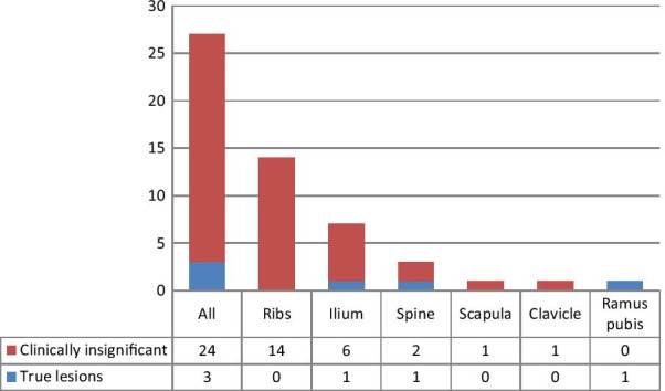
Thirteen of the 15 patients had an overall PSMA-RADS score of 3B, of whom only two had true metastatic disease. The remaining patients had scores of 4 (n = 1) or 5 (n = 1), all confirmed as true positive prostate-related malignant lesions. A per-lesion analysis identified 29 bone lesions, of which 27 were scored PSMA-RADS 3B, and only three of them were true metastases. Thus, debatable lesions proved to have no clinical significance in 84.6% of cases, and only 11% of equivocal PSMA-RADS 3B bone lesions were true positive.

CONCLUSIONS

In intermediate and high-risk patients staged prior to radical prostatectomy, the majority of PSMA-RADS 3B lesions are of no clinical relevance. Bone lesions judged as being highly suspicious for metastases (PSMA-RADS 4/5) were all validated as true positives.

摘要

背景

评估新诊断前列腺癌(PC)患者的疾病范围对于制定合适的治疗方案至关重要。据报道,前列腺特异性膜抗原(PSMA)靶向正电子发射断层扫描/计算机断层扫描(PET/CT)在PC分期方面比传统成像具有更高的准确性。与任何成像方式一样,也会出现陷阱和非特异性发现。PSMA报告和数据系统(PSMA-RADS)1.0版提供了对PSMA靶向研究的结构化解读,并根据临床意义的可能性对病变进行分类。这项回顾性研究的目的是评估术前PSMA靶向成像中,根据PSMA-RADS 1.0版定义的可疑骨病变的临床意义。在406例接受根治性前列腺切除术前行PET/CT分期的连续患者中,有15例存在可疑骨病变。对扫描结果采用PSMA-RADS 1.0系统进行回顾性评分,对疾病进程和随访数据进行盲法处理。影像学和组织学检查结果支持的术后前列腺特异性抗原水平持续存在,被用作可疑影像学检查结果真正意义的参考标准。

结果

15例患者中有13例的PSMA-RADS总评分为3B,其中只有2例有真正的转移性疾病。其余患者评分为4分(n = 1)或5分(n = 1),均被确认为真正的前列腺相关恶性阳性病变。逐病变分析确定了29个骨病变,其中27个评分为PSMA-RADS 3B,其中只有3个是真正的转移灶。因此,在84.6%的病例中,可疑病变被证明无临床意义,PSMA-RADS 3B的可疑骨病变中只有11%为真正的阳性。

结论

在根治性前列腺切除术前行分期的中高危患者中,大多数PSMA-RADS 3B病变无临床相关性。被判定为高度怀疑转移的骨病变(PSMA-RADS 4/5)均被确认为真正的阳性。

https://cdn.ncbi.nlm.nih.gov/pmc/blobs/9955/7788112/b3210303ca79/13550_2020_745_Fig3_HTML.jpg
https://cdn.ncbi.nlm.nih.gov/pmc/blobs/9955/7788112/34bdae45a98b/13550_2020_745_Fig1_HTML.jpg
https://cdn.ncbi.nlm.nih.gov/pmc/blobs/9955/7788112/6176209529b5/13550_2020_745_Fig2_HTML.jpg
https://cdn.ncbi.nlm.nih.gov/pmc/blobs/9955/7788112/b3210303ca79/13550_2020_745_Fig3_HTML.jpg
https://cdn.ncbi.nlm.nih.gov/pmc/blobs/9955/7788112/34bdae45a98b/13550_2020_745_Fig1_HTML.jpg
https://cdn.ncbi.nlm.nih.gov/pmc/blobs/9955/7788112/6176209529b5/13550_2020_745_Fig2_HTML.jpg
https://cdn.ncbi.nlm.nih.gov/pmc/blobs/9955/7788112/b3210303ca79/13550_2020_745_Fig3_HTML.jpg

相似文献

1
The significance of equivocal bone findings in staging PSMA imaging in the preoperative setting: validation of the PSMA-RADS version 1.0.术前PSMA成像分期中骨显像结果不明确的意义:PSMA-RADS 1.0版的验证
EJNMMI Res. 2021 Jan 6;11(1):3. doi: 10.1186/s13550-020-00745-8.
2
[Ga]Ga-PSMA-11 PET and Prostate Cancer Bone Metastases: Diagnostic Performance of Available Standardized Criteria.镓-PSMA-11 PET 与前列腺癌骨转移:现有标准化标准的诊断性能。
J Nucl Med. 2024 Sep 3;65(9):1376-1382. doi: 10.2967/jnumed.124.267899.
3
Follow-up of Lesions with Equivocal Radiotracer Uptake on PSMA-Targeted PET in Patients with Prostate Cancer: Predictive Values of the PSMA-RADS-3A and PSMA-RADS-3B Categories.前列腺癌患者 PSMA 靶向 PET 放射性示踪剂摄取可疑病灶的随访:PSMA-RADS-3A 和 PSMA-RADS-3B 类别的预测值。
J Nucl Med. 2019 Apr;60(4):511-516. doi: 10.2967/jnumed.118.217653. Epub 2018 Sep 6.
4
More advantages in detecting bone and soft tissue metastases from prostate cancer using F-PSMA PET/CT.使用F-PSMA PET/CT检测前列腺癌骨和软组织转移方面有更多优势。
Hell J Nucl Med. 2019 Jan-Apr;22(1):6-9. doi: 10.1967/s002449910952. Epub 2019 Mar 7.
5
Indeterminate skeletal and lymph node lesion on 18F PSMA 1007 PET/CT scanning: lessons from a review at 12 months with PSMA-RADS.18F PSMA 1007 PET/CT 扫描发现不确定的骨骼和淋巴结病变:PSMA-RADS 12 个月复查的经验教训。
Nucl Med Commun. 2022 Sep 1;43(9):1034-1041. doi: 10.1097/MNM.0000000000001600. Epub 2022 Jul 14.
6
Interobserver agreement of [Ga]Ga-PSMA-11 PET/CT images interpretation in men with newly diagnosed prostate cancer.[镓]Ga-PSMA-11 PET/CT图像解读在新诊断前列腺癌男性患者中的观察者间一致性。
EJNMMI Res. 2020 Feb 28;10(1):15. doi: 10.1186/s13550-020-0596-4.
7
Effect of Point-Spread Function Reconstruction for Indeterminate PSMA-RADS-3A Lesions on PSMA-Targeted PET Imaging of Men with Prostate Cancer.点扩散函数重建对前列腺癌男性患者PSMA靶向PET成像中不确定的PSMA-RADS-3A病变的影响。
Diagnostics (Basel). 2021 Apr 7;11(4):665. doi: 10.3390/diagnostics11040665.
8
Ga-Labeled Prostate-specific Membrane Antigen Ligand Positron Emission Tomography/Computed Tomography for Prostate Cancer: A Systematic Review and Meta-analysis.Ga 标记前列腺特异性膜抗原配体正电子发射断层扫描/计算机断层扫描在前列腺癌中的应用:系统评价和荟萃分析。
Eur Urol Focus. 2018 Sep;4(5):686-693. doi: 10.1016/j.euf.2016.11.002. Epub 2016 Nov 15.
9
Changing Threshold-Based Segmentation Has No Relevant Impact on Semi-Quantification in the Context of Structured Reporting for PSMA-PET/CT.在PSMA-PET/CT结构化报告的背景下,改变基于阈值的分割对半定量分析没有显著影响。
Cancers (Basel). 2022 Jan 6;14(2):270. doi: 10.3390/cancers14020270.
10
Recent Updates on Molecular Imaging Reporting and Data Systems (MI-RADS) for Theranostic Radiotracers-Navigating Pitfalls of SSTR- and PSMA-Targeted PET/CT.用于治疗诊断放射性示踪剂的分子影像报告和数据系统(MI-RADS)的最新进展——应对生长抑素受体(SSTR)和前列腺特异性膜抗原(PSMA)靶向PET/CT的陷阱
J Clin Med. 2019 Jul 19;8(7):1060. doi: 10.3390/jcm8071060.

引用本文的文献

1
Interobserver and intraobserver agreement for equivocal lesions in Ga-68 PSMA PET/CT according to PSMA-RADS 2.0 criteria.根据PSMA-RADS 2.0标准,68Ga-PSMA PET/CT中可疑病变的观察者间和观察者内一致性。
Ann Nucl Med. 2025 Sep 10. doi: 10.1007/s12149-025-02103-x.
2
Comparison of Prostate-specific Membrane Antigen Reporting and Data System Version 1.0 versus 2.0 for Prostate Cancer Assessment.用于前列腺癌评估的前列腺特异性膜抗原报告与数据系统1.0版和2.0版的比较
Radiol Imaging Cancer. 2025 May;7(3):e240390. doi: 10.1148/rycan.240390.
3
Structured reporting in prostate cancer: the revolution of quality in nuclear medicine scan interpretation.

本文引用的文献

1
False positive PSMA PET for tumor remnants in the irradiated prostate and other interpretation pitfalls in a prospective multi-center trial.阳性前列腺特异性膜抗原 PET 预测前列腺癌根治术后肿瘤残留:一项前瞻性多中心研究
Eur J Nucl Med Mol Imaging. 2021 Feb;48(2):501-508. doi: 10.1007/s00259-020-04945-1. Epub 2020 Aug 17.
2
Interobserver and intraobserver agreement on prostate-specific membrane antigen PET/CT images according to the miTNM and PSMA-RADS criteria.基于 miTNM 和 PSMA-RADS 标准的前列腺特异性膜抗原 PET/CT 图像的观察者间和观察者内一致性。
Nucl Med Commun. 2020 Aug;41(8):759-767. doi: 10.1097/MNM.0000000000001219.
3
前列腺癌的结构化报告:核医学扫描解读质量的变革。
Eur Radiol. 2024 Feb;34(2):1155-1156. doi: 10.1007/s00330-023-10507-4. Epub 2023 Dec 20.
4
Medicinal (Radio) Chemistry: Building Radiopharmaceuticals for the Future.药用(放射性)化学:为未来构建放射性药物。
Curr Med Chem. 2024;31(34):5481-5534. doi: 10.2174/0929867331666230818092634.
5
PSMA PET imaging in the diagnosis and management of prostate cancer.PSMA PET 成像在前列腺癌的诊断和治疗中的应用。
Abdom Radiol (NY). 2023 Dec;48(12):3610-3623. doi: 10.1007/s00261-023-04002-z. Epub 2023 Jul 26.
6
Prostate-specific Membrane Antigen Reporting and Data System Version 2.0.前列腺特异性膜抗原报告和数据系统第 2.0 版。
Eur Urol. 2023 Nov;84(5):491-502. doi: 10.1016/j.eururo.2023.06.008. Epub 2023 Jul 4.
7
PET Criteria by Cancer Type from Imaging Interpretation to Treatment Response Assessment: Beyond FDG PET Score.从影像解读到治疗反应评估的按癌症类型划分的PET标准:超越FDG PET评分
Life (Basel). 2023 Feb 22;13(3):611. doi: 10.3390/life13030611.
8
Predicting Outcomes of Indeterminate Bone Lesions on F-DCFPyL PSMA PET/CT Scans in the Setting of High-Risk Primary or Recurrent Prostate Cancer.预测高风险原发性或复发性前列腺癌患者 F-DCFPyL PSMA PET/CT 扫描中不确定骨病变的结果。
J Nucl Med. 2023 Mar;64(3):395-401. doi: 10.2967/jnumed.122.264334. Epub 2022 Oct 20.
9
Assessment of malignancy and PSMA expression of uncertain bone foci in [F]PSMA-1007 PET/CT for prostate cancer-a single-centre experience of PET-guided biopsies.评估 [F]PSMA-1007 PET/CT 中前列腺癌不确定骨病灶的恶性程度和 PSMA 表达——一项 PET 引导活检的单中心经验。
Eur J Nucl Med Mol Imaging. 2022 Sep;49(11):3910-3916. doi: 10.1007/s00259-022-05745-5. Epub 2022 Apr 28.
10
Freely available artificial intelligence for pelvic lymph node metastases in PSMA PET-CT that performs on par with nuclear medicine physicians.可自由获取的用于 PSMA PET-CT 中盆腔淋巴结转移的人工智能,其表现与核医学医师相当。
Eur J Nucl Med Mol Imaging. 2022 Aug;49(10):3412-3418. doi: 10.1007/s00259-022-05806-9. Epub 2022 Apr 27.
Factors Predicting Metastatic Disease in Ga-PSMA-11 PET-Positive Osseous Lesions in Prostate Cancer.
预测前列腺癌 Ga-PSMA-11 PET 阳性骨病变转移疾病的因素。
J Nucl Med. 2020 Dec;61(12):1779-1785. doi: 10.2967/jnumed.119.241174. Epub 2020 Apr 17.
4
Prostate-specific membrane antigen PET-CT in patients with high-risk prostate cancer before curative-intent surgery or radiotherapy (proPSMA): a prospective, randomised, multicentre study.高风险前列腺癌患者在接受根治性手术或放疗前的前列腺特异性膜抗原 PET-CT(proPSMA):一项前瞻性、随机、多中心研究。
Lancet. 2020 Apr 11;395(10231):1208-1216. doi: 10.1016/S0140-6736(20)30314-7. Epub 2020 Mar 22.
5
Intraindividual Comparison of F-PSMA-1007 with Renally Excreted PSMA Ligands for PSMA PET Imaging in Patients with Relapsed Prostate Cancer.个体内比较 F-PSMA-1007 与肾排泄 PSMA 配体在复发性前列腺癌患者 PSMA PET 成像中的应用。
J Nucl Med. 2020 May;61(5):729-734. doi: 10.2967/jnumed.119.234898. Epub 2019 Oct 18.
6
Head-to-Head Comparison of Ga-PSMA-11 with F-PSMA-1007 PET/CT in Staging Prostate Cancer Using Histopathology and Immunohistochemical Analysis as a Reference Standard.头对头比较 Ga-PSMA-11 与 F-PSMA-1007 PET/CT 在前列腺癌分期中使用组织病理学和免疫组织化学分析作为参考标准。
J Nucl Med. 2020 Apr;61(4):527-532. doi: 10.2967/jnumed.119.234187. Epub 2019 Sep 27.
7
Matched-Pair Comparison of Ga-PSMA-11 PET/CT and F-PSMA-1007 PET/CT: Frequency of Pitfalls and Detection Efficacy in Biochemical Recurrence After Radical Prostatectomy.镓-PSMA-11 PET/CT 与 F-PSMA-1007 PET/CT 的配对比较:根治性前列腺切除术后生化复发时的陷阱频率和检测效能。
J Nucl Med. 2020 Jan;61(1):51-57. doi: 10.2967/jnumed.119.229187. Epub 2019 Jun 28.
8
Prostate Cancer, Version 2.2019, NCCN Clinical Practice Guidelines in Oncology.《前列腺癌(2019 年版)》,NCCN 肿瘤学临床实践指南。
J Natl Compr Canc Netw. 2019 May 1;17(5):479-505. doi: 10.6004/jnccn.2019.0023.
9
Assessment of 68Ga-PSMA-11 PET Accuracy in Localizing Recurrent Prostate Cancer: A Prospective Single-Arm Clinical Trial.68Ga-PSMA-11 PET 定位复发性前列腺癌的准确性评估:一项前瞻性单臂临床试验。
JAMA Oncol. 2019 Jun 1;5(6):856-863. doi: 10.1001/jamaoncol.2019.0096.
10
Persistent Prostate-Specific Antigen After Radical Prostatectomy and Its Impact on Oncologic Outcomes.根治性前列腺切除术后前列腺特异性抗原持续存在及其对肿瘤学结局的影响。
Eur Urol. 2019 Jul;76(1):106-114. doi: 10.1016/j.eururo.2019.01.048. Epub 2019 Feb 14.